

Case Reports
doi: 10.1002/ccr3.1999.
eCollection 2019 Mar.
Dermoscopic clues in the skin lesions of secondary syphilis
Affiliations
- PMID: 30899465
- PMCID: PMC6406165
- DOI: 10.1002/ccr3.1999
Item in Clipboard
Case Reports
Dermoscopic clues in the skin lesions of secondary syphilis
Clin Case Rep.
.
Abstract
Secondary syphilis may have a varied clinical presentation and might pose a diagnostic difficulty when a typical history is absent. We describe the dermoscopic clues of the skin lesions at different stages of the disease which could culminate to a proper diagnosis.
Keywords: Biett’s sign; dermatoscopy; dermoscopy; infectious disease; syphilis.
Conflict of interest statement
None declared.
Figures
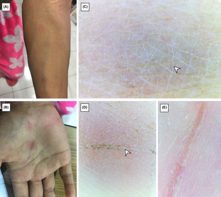
A and B, Clinically, erythematous maculopapular rash can be seen over left forearm and right palm. Dermoscopy (polarized dermoscopy, original magnification × 20) of lesions over forearm, C and palm, D showing scaling within the skin furrows (white arrowheads) and central darker area fading toward the periphery with an ill‐defined border. Orange color of the lesion can be noted in the lesion over palm. E, Dermoscopy (polarized dermoscopy, original magnification ×20) of a psoriatic lesion over palm from a different patient showing larger scales, pinkish color and dotted vessels
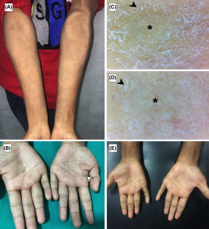
A and B, Clinically, lesions with increased scaling and decreased erythema can be seen over bilateral forearms and palms, respectively, on the 5th day of initial presentation. Peripheral scaling can be clearly noted in the lesions over palms. C and D, Dermoscopy (polarized dermoscopy, original magnification × 20) of lesions over forearm during the visit showing peripheral scaling with a relatively clear central area. E, Clearance of scaling seen on palms on the 14th day of treatment
Similar articles
-
Syphilis, the Great Imitator-Clinical and Dermoscopic Features of a Rare Presentation of Secondary Syphilis.Int J Environ Res Public Health. 2023 Jan 11;20(2):1339. doi: 10.3390/ijerph20021339. Int J Environ Res Public Health. 2023. PMID: 36674095 Free PMC article.
-
Unusual Clinical Presentation of Giant Extragenital Condyloma.Acta Dermatovenerol Croat. 2020 Dec;28(7):240-241. Acta Dermatovenerol Croat. 2020. PMID: 33834999
-
Comment on: "Dermoscopy of Biett's sign and differential diagnosis with annular maculopapular rashes with scaling".Indian J Dermatol Venereol Leprol. 2018 Jul-Aug;84(4):441-442. doi: 10.4103/ijdvl.IJDVL_97_18. Indian J Dermatol Venereol Leprol. 2018. PMID: 29893299 No abstract available.
-
Dermatoscopy of Inflammatory Diseases in Skin of Color.Indian Dermatol Online J. 2021 Jan 16;12(1):45-57. doi: 10.4103/idoj.IDOJ_613_20. eCollection 2021 Jan-Feb. Indian Dermatol Online J. 2021. PMID: 33768022 Free PMC article. Review.
-
Dermoscopy for venereologists: an update on patterns of tumors, inflammatory and infectious diseases of the genitalia, and tips for differential diagnosis.Int J Dermatol. 2021 Oct;60(10):1211-1218. doi: 10.1111/ijd.15333. Epub 2020 Dec 8. Int J Dermatol. 2021. PMID: 33448049 Review.
Cited by
-
Dermoscopy of skin infestations and infections (entomodermoscopy) - Part I: dermatozoonoses and bacterial infections.An Bras Dermatol. 2021 Nov-Dec;96(6):735-745. doi: 10.1016/j.abd.2021.04.007. Epub 2021 Oct 5. An Bras Dermatol. 2021. PMID: 34620524 Free PMC article. Review.
-
Secondary Syphilis Presents as Palmoplantar Hyperpigmented Maculopapules: A Case Report.Cureus. 2024 Apr 1;16(4):e57367. doi: 10.7759/cureus.57367. eCollection 2024 Apr. Cureus. 2024. PMID: 38566778 Free PMC article.
-
From clinical clues to histologic truth: A case of condyloma lata.Indian J Sex Transm Dis AIDS. 2025 Jan-Jun;46(1):88-89. doi: 10.4103/ijstd.ijstd_142_24. Epub 2025 Jun 9. Indian J Sex Transm Dis AIDS. 2025. PMID: 40546362 Free PMC article. No abstract available.
-
Syphilis, the Great Imitator-Clinical and Dermoscopic Features of a Rare Presentation of Secondary Syphilis.Int J Environ Res Public Health. 2023 Jan 11;20(2):1339. doi: 10.3390/ijerph20021339. Int J Environ Res Public Health. 2023. PMID: 36674095 Free PMC article.
-
Dermoscopic Features and Their Diagnostic Values Among Common Inflammatory and Infectious Dermatoses: A Cross-Sectional Study.Clin Cosmet Investig Dermatol. 2023 Jan 24;16:211-220. doi: 10.2147/CCID.S397212. eCollection 2023. Clin Cosmet Investig Dermatol. 2023. PMID: 36718215 Free PMC article.
References
-
- Peeling RW, Hook EW. The pathogenesis of syphilis: the Great Mimicker, revisited. J Pathol. 2006;208:224–232. - PubMed
-
- Angus J, Langan SM, Stanway A, Leach IH, Littlewood SM, English JS. The many faces of secondary syphilis:a re‐emergence of an old disease. Clin Exp Dermatol. 2006;31:741–745. - PubMed
-
- Errichetti E, Stinco G. Dermoscopy in differentiating palmar syphiloderm from palmar papular psoriasis. Int J STD AIDS. 2017;28:1461–1463. - PubMed
-
- Tognetti L, Sbano P, Fimiani M, Rubegni P. Dermoscopy of Biett's sign and differential diagnosis with annular maculo‐papular rashes with scaling. Indian J Dermatol Venereol Leprol. 2017;83:270–273. - PubMed
-
- Martín IG, Ankad BS, Errichetti E, Lallas A, Ioannides D, Zaballos P. Bacterial and parasitic infections In: Lallas A, Errichetti E, Ioannides D, eds. Dermoscopy in General Dermatology. Boca Raton: CRC Press; 2018:190–192.
Publication types
LinkOut - more resources
Full Text Sources
