

doi: 10.1016/j.eats.2018.09.006.
eCollection 2019 Jan.
Distal Triceps Rupture Repair: The Triceps Pulley-Pullover Technique
Affiliations
- PMID: 30899656
- PMCID: PMC6408749
- DOI: 10.1016/j.eats.2018.09.006
Item in Clipboard
Distal Triceps Rupture Repair: The Triceps Pulley-Pullover Technique
Arthrosc Tech.
.
Abstract
Distal triceps rupture is an uncommon but debilitating injury, and surgical fixation is almost invariably warranted. A number of techniques have been described in the literature in which combinations of transosseous tunnels and bone anchors have been used. We describe a modification to existing techniques-the triceps pulley-pullover technique with all-suture anchors. This technique minimizes bone loss, while maximizing the bone-tendon contact area and creating a double-row repair to optimize strength and healing.
Figures

The patient is placed in the lateral decubitus position with the operative arm (left in this case) over a bar.

A standard posterior approach to the distal triceps and proximal 5 to 8 cm of ulna is used to expose the distal triceps tendon. Patient in lateral decubitus position.

The triceps tendon is debrided and the footprint identified and prepared with a curette.

This sketch shows the preparation of the triceps tendon footprint. A curette is used to debride the bone to a fresh bleeding surface to optimize healing of the tendon to the bone.

Two all-suture anchors are inserted into the triceps footprint. Here the anchor on the radial side (anchor AB) has already been inserted and the anchor on the ulna side (anchor CD) is now being inserted by tapping it into a predrilled 2.3-mm–diameter pilot hole. The anchors are positioned approximately 15 mm from the ulna tip and 15 mm apart.

The 2 all-suture 2.3-mm anchors are inserted (labeled AB and CD), approximately 15 mm apart, to form the proximal row. Note that anchor AB includes sutures A and B; anchor CD includes sutures C and D.

The all-suture anchors shown in cross-section through the proximal ulna: anchor AB on the lateral (radial) side of the footprint and anchor CD on the medial (ulna) side of the footprint. Here each anchor contains 4 strands (2 lengths of suture); however, anchors with more sutures may be used.

Forming the pulley. The A1 and C1 strands are passed from deep to superficial through the distal triceps tendon; this is repeated for the A2 and C2 strands 5 to 10 mm distal to the first pair. Here a loop-stitch is being used as a suture passer, and 6 strand anchors have been used.
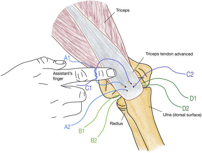
Forming the pulley. After the A and C strands have been passed through the triceps tendon from deep to superficial (orange arrows), strands A1 and C1 are tied over the assistant's finger to form a loop. Note B and D strands still lie under the distal triceps tendon.

The pulley technique. Pulling on A2 and C2 (long orange arrows) draws the knot formed by A1 and C1, advancing and snugging the tendon down to the footprint (short orange arrows).

The pulley technique. A2 is tied to C2 over the triceps tendon, thus locking the pulley strands in place.

Augmentation. Strands B1/B2 and D1/D2 are used to form locking stitches along the medial and lateral borders of the triceps. A Krakow technique or a similar technique can be used.

The pullover technique. Pulling the locked strands distally over the tendon augments and braces the repair. Here the site of the distal anchor is being planned.

The pullover technique. Pulling the strands from the knots distally tensions and advances the distal triceps over the original footprint. In this example, 4 strands have been used, but more can be taken distally, depending on the capacity of the push-lock anchor chosen by the surgeon for the distal row.
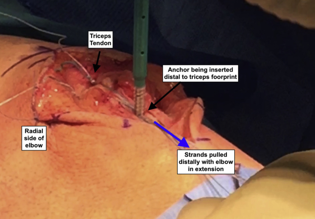
The pullover technique. Placement of the distal row locking anchor in the dorsal border of the ulna, distal to the triceps insertion. This must be done with the elbow in extension.

The pullover technique. The distal row suture anchor is placed using strands A1, B1, C1 and D1 forming the distal second row. Remaining strands are cut short.

The distal row suture anchor in situ to complete the repair. This must be locked down with the elbow in extension. In this case 6 strands have been, advanced from the knots of the proximal row; however, 4 strands would be adequate. The remaining strands are cut short.
References
-
- Anzel S.H., Covey K.W., Weiner A.D., Lipscomb P.R. Disruption of muscles and tendons: An analysis of 1,014 cases. Surgery. 1959;45:406–414. - PubMed
-
- Horneff J.G., 3rd, Aleem A., Nicholson T. Functional outcomes of distal triceps tendon repair comparing transosseous bone tunnels with suture anchor constructs. J Shoulder Elbow Surg. 2017;26:2213–2219. - PubMed
-
- Giannicola G., Bullitta G., Rotini R. Results of primary repair of distal triceps tendon ruptures in a general population: A multicentre study. Bone Joint J. 2018;100:610–616. - PubMed
LinkOut - more resources
Full Text Sources
