. 2019 Jan 21;8(2):e153-e161.
doi: 10.1016/j.eats.2018.10.010. eCollection 2019 Feb.

Anatomic Reconstruction of the Posterolateral Corner: An All-Arthroscopic Technique

Affiliations

Anatomic Reconstruction of the Posterolateral Corner: An All-Arthroscopic Technique

Jannik Frings et al. Arthrosc Tech. .

Abstract

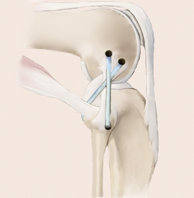
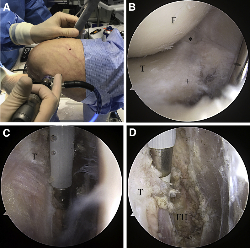
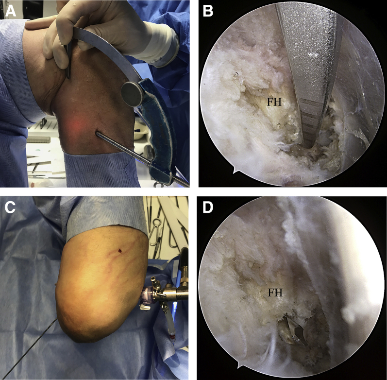
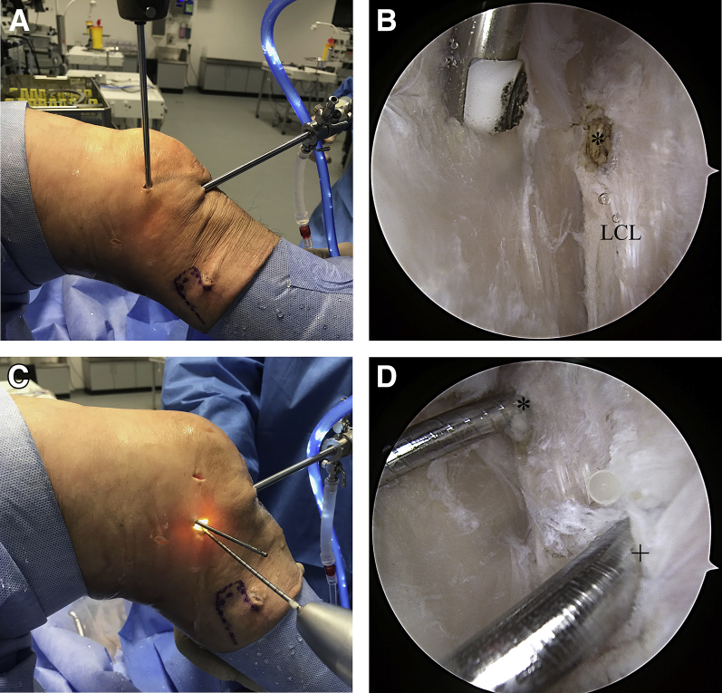
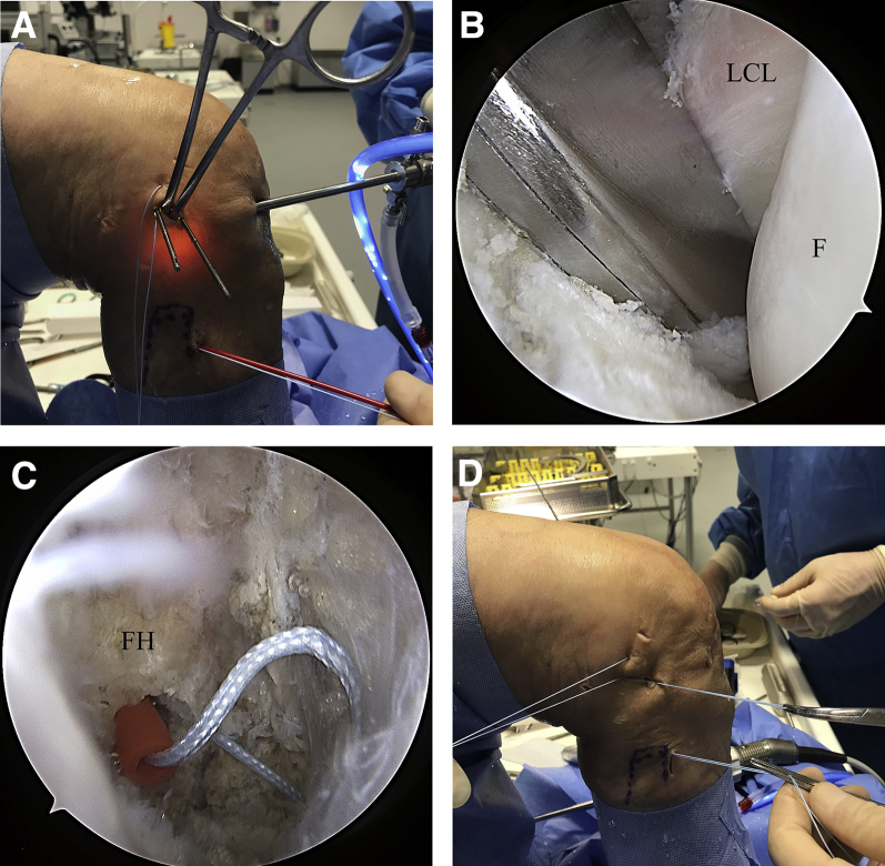
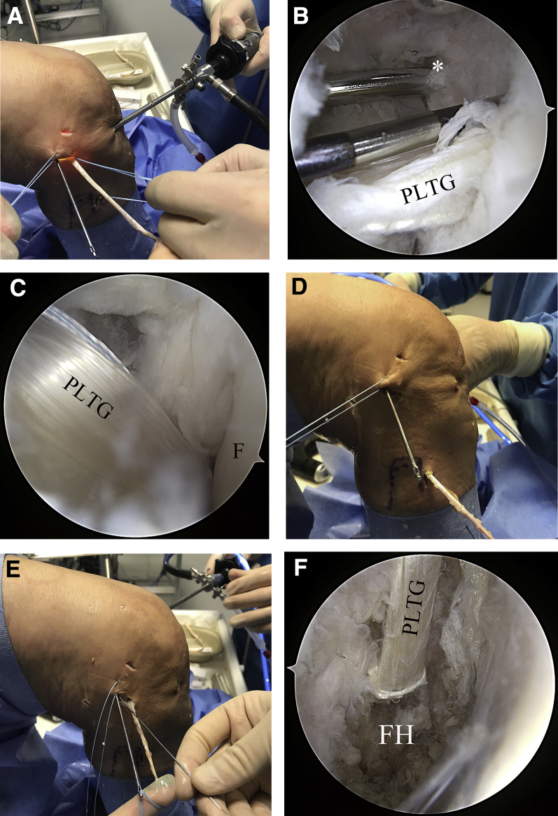
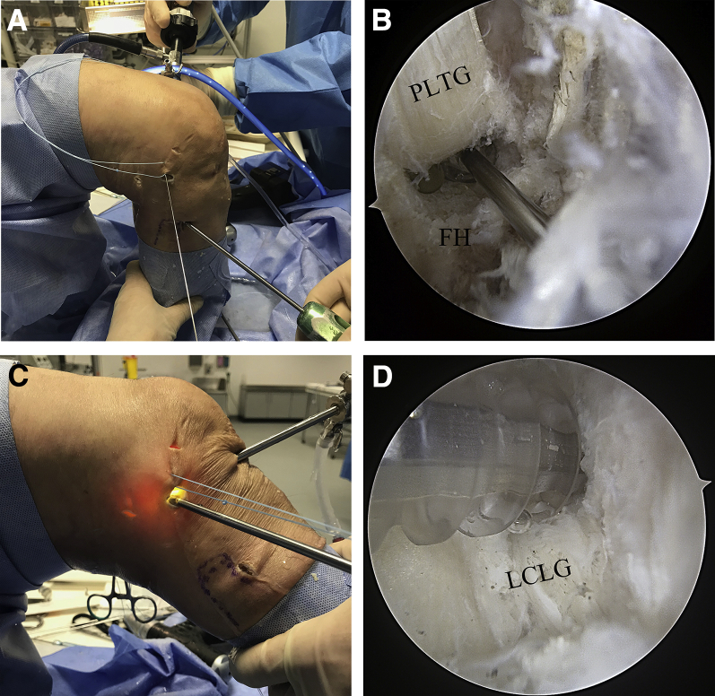
Injuries of the posterolateral corner (PLC) of the knee lead to chronic lateral and external rotational instability. Successful treatment of PLC injuries requires an understanding of the complex anatomy and biomechanics of the PLC. Several open PLC reconstruction techniques have been published. It is understood that anatomic reconstruction is superior to extra-anatomic techniques, leading to better clinical results. An open, anatomic, fibula-based technique for reconstruction to address lateral and rotational instability has been described. However, when an open technique is used, surgeon and patient are faced with disadvantages, such as soft tissue damage or exposure of vulnerable structures. Few arthroscopic techniques for tibia- or fibula-based reconstruction of rotational posterolateral instability have been described. A complete arthroscopic stabilization of the combined lateral and posterolateral rotational instability of the knee has not yet been described. We therefore present the first all-arthroscopic technique for complete PLC reconstruction, based on an open technique described previously. All relevant landmarks of the PLC can be arthroscopically visualized in detail, allowing safe and effective treatment of PLC injuries.


Figures

Fig 1
Fig 1
A fibula-based technique, as described by Arciero, can be used for anatomic reconstruction of the posterolateral corner. The graft is fixed precisely at the anatomic femoral footprints of the LCL and PLT, and the fibular tunnel is drilled in an anterolateral-to-posteromedial direction, to anatomically reconstruct the LCL and PLT footprints. (LCL, lateral collateral ligament; PLT, popliteus muscle tendon.)
Fig 2
Fig 2
For arthroscopic preparation and exposure of the FH (right knee), the patient is placed in the supine position, with the knee secured in an electric leg holder. (A) The dorsolateral recessus can be observed through the posteromedial portal. (B) The posterolateral portal provides access to the capsular fibers (+) posterior to the PLT; these fibers are resected with a shaver (Excalibur, Arthrex) to expose the PLT (*) and the FH. (C) A radiofrequency electrode is used to carefully resect soft tissue until the FH is exposed. (D) Care should be taken to protect the peroneal nerve, which is located an additional 2 to 3 cm distally, as well as the popliteal nerves and vessels. (F, femur; FH, fibular head; PLT, popliteus muscle tendon; T, tibia.)
Fig 3
Fig 3
For preparation of the fibular tunnel (right knee, supine position, arthroscope in posteromedial portal), a tibial drill guide (AR-1510F; Arthrex) is introduced through the posterolateral portal. (A) A small incision is made anteroposterior to the FH, which can be easily palpated. (B) The tip of the drill guide is placed on the dorsomedial surface of the FH, which is visualized arthroscopically through the posteromedial portal. (C) A guidewire is then introduced in an anterolateral-to-posteromedial direction under arthroscopic visualization. (D) The fibular tunnel is drilled with a 5- or 6-mm drill, depending on the diameter of the tendon graft. (FH, fibular head.)
Fig 4
Fig 4
To prepare the femoral tunnels (right knee, supine position, slightly flexed angle), the arthroscope is introduced through the high anterolateral portal, and a parapatellar portal is developed for the shaver. (A) The lateral joint capsule is carefully resected with the shaver (Torpedo; Arthrex) to expose the femoral attachments of the LCL (*) and PLT. The femoral footprints can be marked with a radiofrequency electrode for better orientation. A stab incision is made directly over the femoral attachments. (C) Two guidewires are introduced into the native origins of the LCL (*) and PLT (+). (D) The femoral tunnels are then drilled with a 5- or 6-mm drill, depending on the strength of the graft. The PLT tunnel should have a length of approximately 3 cm, and the LCL tunnel should be of greater depth to allow full graft tensioning. (LCL, lateral collateral ligament; PLT, popliteus muscle tendon.)
Fig 5
Fig 5
Anatomic graft positioning requires an exact implementation of the shuttling sutures, following the course of the native PLT (right knee, supine position). (A) To obtain a better overview, stay sutures can be used to retract the iliotibial band. (B) A bent clamp is introduced through the lateral stab incision and is then passed down along the popliteus tendon. This step should be observed arthroscopically to ensure correct placement. (C) A FiberStick is inserted through the fibular tunnel, and the FiberWire is passed into the posterolateral recessus, where it is grasped with the bent clamp. (D) The FiberWire is then shuttled through the lateral stab incision and the fibular tunnel. To provide more space for the clamp, the femoral guidewires may be retracted so that the tips slightly overtop the lateral cortex. (PLT, popliteus muscle tendon.)
Fig 6
Fig 6
We recommend using a gracilis or semitendinosus tendon graft, with a length of at least 20 cm. (A) The armed graft is shuttled into the femoral PLT tunnel and fixed with a bioabsorbable interference screw. (B) Again, a nitinol wire may be of use for retaining the tunnel position while the PLTG is fixed; the LCL (*) drill channel can be seen posterior to the PLT tunnel. (C) The PLTG is then shuttled into the posterolateral recessus, along the native PLT. At this point, precise anatomic placement of the graft is crucial. Interposition of soft tissue must be meticulously avoided. (D) The graft is further shuttled through the fibular tunnel in the posteromedial-to-anterolateral direction. (E) The graft is then shuttled to the lateral stab incision and into the femoral LCL tunnel. (F) The implanted PLTG can be observed though the posteromedial portal. (LCL, lateral collateral ligament; PLT, popliteus muscle tendon; PLTG, popliteus tendon graft.)
Fig 7
Fig 7
(A) During fibular graft fixation (right knee, supine position), the knee is bent to 90°, and the graft is carefully tightened. (B) A bioabsorbable interference screw is introduced along a nitinol wire into the fibular tunnel. Correct screw placement can be verified under arthroscopic visualization. (C) After PLT fixation, the graft is fixed in the LCL tunnel while the knee is bent to 20° to 30° of flexion. (D) During fixation with a bioabsorbable interference screw, overtopping of the screws must be prevented by means of arthroscopic visualization to avoid painful soft tissue irritation. (LCL, lateral collateral ligament; PLT, popliteus muscle tendon.)

References

    1. LaPrade R.F., Griffith C.J., Coobs B.R., Geeslin A.G., Johansen S., Engebretsen L. Improving outcomes for posterolateral knee injuries. J Orthop Res. 2014;32:485–491. - PubMed
    1. Frosch K.-H., Akoto R., Heitmann M., Enderle E., Giannakos A., Preiss A. Arthroscopic reconstruction of the popliteus complex: Accuracy and reproducibility of a new surgical technique. Knee Surg Sports Traumatol Arthrosc. 2015;23:3114–3120. - PubMed
    1. Thaunat M., Pioger C., Chatellard R., Conteduca J., Khaleel A., Sonnery-Cottet B. The arcuate ligament revisited: Role of the posterolateral structures in providing static stability in the knee joint. Knee Surg Sports Traumatol Arthrosc. 2014;22:2121–2127. - PubMed
    1. Shon O.-J., Park J.-W., Kim B.-J. Current concepts of posterolateral corner injuries of the knee. Knee Surg Relat Res. 2017;29:256. - PMC - PubMed
    1. Domnick C., Frosch K.H., Raschke M.J. Kinematics of different components of the posterolateral corner of the knee in the lateral collateral ligament-intact state: A human cadaveric study. Arthroscopy. 2017;33:1821–1830. - PubMed