Review
. 2019 Mar 21;10(1):38.
doi: 10.1186/s13244-019-0716-8.

All about portal vein: a pictorial display to anatomy, variants and physiopathology

Affiliations
Review

All about portal vein: a pictorial display to anatomy, variants and physiopathology

Carolina Carneiro et al. Insights Imaging. .

Abstract

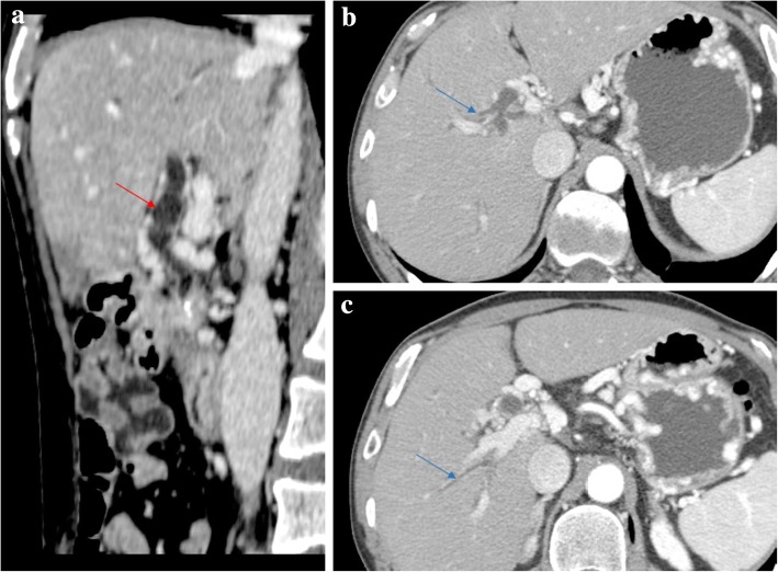
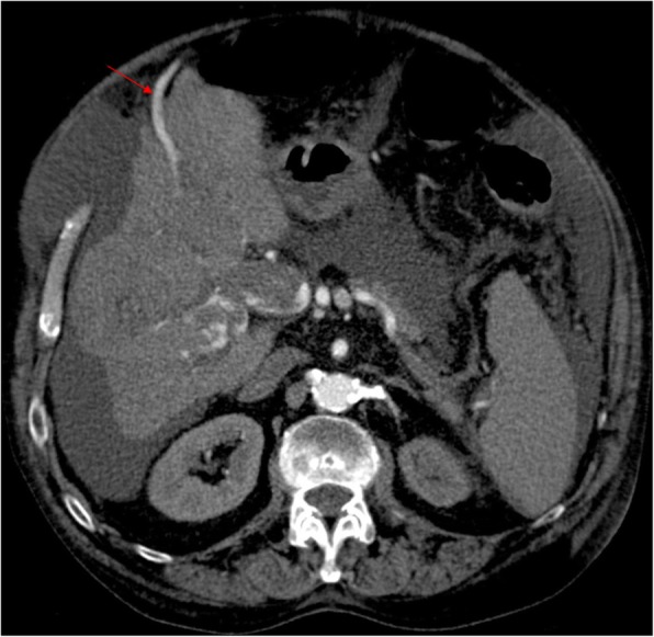
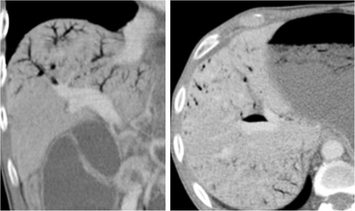
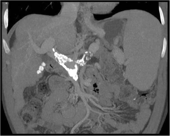
The portal vein (PV) is the main vessel of the portal venous system (PVS), which drains the blood from the gastrointestinal tract, gallbladder, pancreas, and spleen to the liver. There are several variants affecting the PV, and quite a number of congenital and acquired pathologies.In this pictorial review, we assess the embryological development and normal anatomy of the PVS, displaying selected cases consisting of normal variants, congenital anomalies, and a large and heterogeneous group of acquired conditions that may affect the PV.

Keywords: Anatomic variation; Portal hypertension; Portal system; Portal vein; Venous thrombosis.


Conflict of interest statement

Competing interests

The authors declare that they have no competing interests.

Publisher’s Note

Springer Nature remains neutral with regard to jurisdictional claims in published maps and institutional affiliations.

Figures

Fig. 1
Fig. 1
Diagrammatic representation of the embryological development of the PV. a The vitelline venous system arrives at the primitive liver as two paired veins (right and left), branches into the hepatic sinusoids, and then coalesce, pierce the septum tranversum (primitive diaphragm) and drain into the sinus venosus (primitive heart). These two vitelline veins communicate through three pre-hepatic anastomoses around the developing duodenum (cranial-ventral, dorsal, and caudal-ventral). b Over time, a selective involution occurs, involving the caudal part of the right vitelline vein, the cranial part of the left vitelline vein, and the caudal-ventral anastomosis. The dorsal and cranial-ventral anastomoses persist and give rise to the main PV and to the left PV, respectively. Initially, the paired umbilical veins lie more lateral than the vitelline ones, and also pierce the septum tranversum and drain into the sinus venosus. With the development of the liver, the umbilical veins fragment and connect to the hepatic sinusoids. Over time, a selective involution of the right umbilical vein and cranial portion of the left umbilical vein also occurs. c The remnant left umbilical vein cranially bifurcates, forming two new communications: one with the IVC through the ductus venosus, carrying oxygenated blood from the placenta directly to the fetus; and another with the left PV, supplying directly the liver. After birth, the ductus venosus and the left umbilical vein involute and become the ligamentum venosum and ligamentum teres, respectively
Fig. 2
Fig. 2
Normal anatomy of the PVS and the typical branching pattern of the main PV. a Volume rendering technique (VRT) image from late arterial phase contrast-enhanced CT depicting a global view of the PVS. b Maximum intensity projection (MIP) image from portal venous phase contrast-enhanced CT showing the typical branching pattern of the main PV
Fig. 3
Fig. 3
Variants of the PV branching pattern. MIP oblique reconstruction images from portal venous phase CT. a PV trifurcation—PV divides into three branches: left PV, right anterior PV, and right posterior PV. b Right posterior PV branch arising directly from the main PV. c Left PV arising from the right anterior segmental branch
Fig. 4
Fig. 4
Preduodenal PV. MPR image from portal venous phase CT shows the main PV coursing in front of the duodenum and pancreas, an incidental finding in this case
Fig. 5
Fig. 5
Circumportal pancreas. Portal venous phase CT axial image shows pancreatic parenchyma surrounding PV like an annulus. In this case, it was a suprasplenic type
Fig. 6
Fig. 6
Congenital intrahepatic portosystemic shunt. MPR image from late arterial phase CT displaying the most common type of congenital intrahepatic portosystemic shunt (type 1), with a single vessel connecting the right PV with the IVC. Note the large caliber of both afferent PV branch and efferent hepatic vein, and the presence of a variceal dilatation between both, appearing as a rounded enhancing mass. If considering this focal varix as an aneurysm, we can classify this shunt as a type 3 either
Fig. 7
Fig. 7
Congenital extrahepatic portosystemic shunt—Abernethy malformation. a VRT image from portal venous phase CT presents a type II Abernethy malformation. SV converges with the SMV giving rise to the main PV. After that, the main PV lacks the classic branching pattern into right and left PV. Instead, a large and dominant aberrant left PV reaches the systemic venous system draining into IVC. Taking into account the origin, the route, and the final confluence with IVC, this aberrant left PV-IVC shunt was interpreted as a patent ductus venosus. b Late arterial phase contrast-enhanced MDCT images demonstrate this was a partial shunting, as we can see small branches arising from the main PV and left PV for both lobes
Fig. 8
Fig. 8
Congenital extrahepatic portosystemic shunt—Abernethy malformation. a VRT image from arterial phase CT of the same patient in Fig. 7. Note aberrant origin of the hepatic artery emerging from superior mesenteric artery. There is also increase caliber of the common hepatic artery and its branches, consequence of the augmented arterial compensatory flow. b Late arterial phase CT images reveal multiple liver nodules (orange arrows), frequently associated with Abernethy malformation [12]
Fig. 9
Fig. 9
Congenital extrahepatic arterioportal fistula. a VRT and b MIP images from dominant arterial phase CT. a Best depict an early enhancement of the PV in this phase, a sign of arterioportal shunt. b Here, we can precisely individualize the arteriovenous malformation, as an aberrant tortuous hepatic artery branch connecting with the left PV, with an attenuation similar to the hepatic artery, filling retrogradely the PV
Fig. 10
Fig. 10
Hereditary hemorrhagic telangiectasia. a Axial fat suppressed T2-weighted MR image shows increased caliber of the hepatic artery and its branches (orange arrows). Note also the enlarged liver with heterogeneous texture. b, c Axial gadolinium-enhanced fat suppressed T1-weighted MR images in the arterial phase show intrahepatic telangiectasias, appearing as rounded small hyperenhancing lesions (b yellow arrow). An early contrast filling of portal venous branches is also seen, both peripheral (b red arrow) and central branches (c blue arrow), in addition to an increased PV caliber
Fig. 11
Fig. 11
Aneurysm of the PVS. a, b Axial and oblique MPR images from late arterial phase CT shows an aneurysmal dilatation of left PV with about 3 cm of greater axis. Note the left PV branches arising directly from the aneurysm (yellow arrow). c VRT from late arterial phase best depict the origin of the aneurysm in the PVS
Fig. 12
Fig. 12
Pseudothrombosis phenomena. a Late arterial phase CT axial image shows a heterogeneous low-attenuation PV content, due to pseudothrombosis phenomena. b Accordingly, on portal venous phase, the PV shows homogeneous fulfilling, discarding a hypothesis of a true thrombosis
Fig. 13
Fig. 13
Acute vs. subacute/chronic PV thrombosis. a Plain CT axial image shows high-attenuation content within a left PV branch (blue arrow)—acute thrombus. b Portal venous phase CT axial image clearly reveals an additional low-attenuation filling defect in the right PV, extending into its right posterior branch (red arrow)—subacute/chronic thrombus
Fig. 14
Fig. 14
Acute portal vein thrombosis. MPR image from portal venous phase CT shows a luminal thrombus within the main PV, and enhancement of the vessel wall (red asterisk), presumably due to still normal flow through its vasa vasorum
Fig. 15
Fig. 15
Portal vein thrombosis, chronic evolution. a MPR and b MIP images from portal venous phase CT show the evolution of a previous left PV complete thrombosis. With time, the unsupplied hepatic segments totally disappeared with atrophic involution of the left lobe
Fig. 16
Fig. 16
Portal vein thrombosis with THED. a Arterial phase CT axial image shows a triangular-shaped area of arterial enhancement, corresponding to the parenchyma formerly supplied by an occluded right anterior PV branch. At portal venous (b) and late phases (c) the hepatic parenchyma homogenizes, and there is persistent absence of venous enhancement due to bland thrombus
Fig. 17
Fig. 17
Cavernomatous transformation of PV. Late arterial phase CT shows absence of a well-defined PV and multiple enhancing veins instead, representing portoportal collateral vessels—portal cavernoma. Note the THED associated with cavernoma
Fig. 18
Fig. 18
Portal biliopathy. Sagittal portal venous phase CT images shows tortuous common bile duct (red arrow) encircled by portal cavernoma, leading to dilation of peripheral bile ducts (blue arrows). Note also gastric varices due to PH
Fig. 19
Fig. 19
Tumoral PV thrombosis. MPR image from early arterial phase CT shows heterogeneous enhancing PV thrombus, despite absent enhancement of both splenic and SMV (not shown). Note also the presence of cirrhosis (favoring hepatocarcinoma as the cause of the thrombus), ascites, and repermeabilization of the paraumbilical vein (red arrow), signs of PH. The early enhancement of the recanalized paraumbilical vein also suggests the presence of malignant arterioportal fistula
Fig. 20
Fig. 20
Portal venous gas. MPR images from portal venous phase CT shows air branching pattern fulfilling some segments of peripheral right PV branches
Fig. 21
Fig. 21
Signs of PH. MIP image showing extensive calcification of PV and its tributaries, in a patient with a long-standing PH. Note the presence of splenomegaly, also a sign of PH, and the incidental finding of gallbladder stones
Fig. 22
Fig. 22
Pre-sinusoidal causes of PH. a MIP image from portal venous phase CT shows extrinsic compression of PV by a large hepatic cyst (red arrow head), a benign cause of pre-sinusoidal PH. b MPR image from portal venous phase CT show PV invasion by a pancreatic adenocarcinoma, a malignant cause of pre-sinusoidal PH
Fig. 23
Fig. 23
Obliterative portal venopathy. 32-year-old female, with HIV infection and slightly increased bilirubin levels, who complained of increased abdominal volume. Axial (a) plain, (b) arterial, and (c) portal venous phase contrast-enhanced CT images show some morphologic liver changes, with hypertrophy of the caudate lobe, but regular contours and no atrophy of segment IV. Right PV is hypoattenuating before and after contrast material administration (yellow arrows), a feature suggesting non-acute thrombus. Hepatic parenchyma displays heterogeneous peripheral enhancement in arterial phase (b) due to compensatory increased arterial flow. This THED fades in portal venous phase (c). Some PH stigmata are present, as such splenomegaly, permeable umbilical vein (red arrow), and other varices. Liver biopsy was performed and OPV diagnosis pathologically confirmed
Fig. 24
Fig. 24
Esophageal and paraesophageal varices. Portal venous phase CT axial images show paraesophageal varices (red arrow) in the mediastinum, appearing as dilated tortuous veins surrounding the outer surface of the esophageal inferior third (green arrow), and esophageal varices (blue arrow), presenting as submucosal varicose veins within the esophagus wall
Fig. 25
Fig. 25
Gastrorenal shunt. MPR image from portal venous phase CT at the level of the left kidney shows a tortuous and high-caliber gastrorenal shunt, running from gastric varices to the left renal vein. Note the increased diameter of the left renal vein downstream shunt confluence, compared to the normal sized left renal vein segment upstream
Fig. 26
Fig. 26
Anorectal/hemorrhoidal varices. Portal venous phase CT images showing a variceal network along, around and within the anorectal wall. In this case, we can individualize ingurgitated tortuous vessels (red arrows), but sometimes thickening and hyperenhancement are all we can see
Fig. 27
Fig. 27
Retroperitoneal varices. VRT image from portal venous phase CT shows an increase in size of SV (green arrow) and SMV (red arrow), an extensive collateral network of varices fed by SMV (blue arrows), communicating with a huge-caliber right ovarian vein (yellow arrow), and giving rise to varices along the ovarian venous plexus
Fig. 28
Fig. 28
Transjugular intrahepatic portosystemic shunt. MIP image from portal venous phase CT shows a metallic stent connecting the PV with the hepatic vein. Note enlargement of the hepatic vein due to increased flow directly drained from the PVS to its lumen, skipping the hepatic parenchyma
Fig. 29
Fig. 29
Liver surgery. MIP image from portal venous phase CT shows right hepatectomy findings: section and ligation of the right PV at its origin; shift of left liver toward the right with horizontalization of PV; prominent caliber of left PV and hypertrophy and rounded contours of the remaining liver

References

    1. Corness JA, McHugh K, Roebuck DJ, Taylor AM. The portal vein in children: radiological review of congenital anomalies and acquired abnormalities. Pediatr Radiol. 2006;36:87–96. doi: 10.1007/s00247-005-0010-4. - DOI - PubMed
    1. Lee WK, Chang SD, Duddalwar VA et al (2011) Imaging assessment of congenital and acquired abnormalities of the portal venous system. Radiographics 31:905–926 - PubMed
    1. Tirumani SH, Shanbhogue AKP, Vikram R, Prasad SR, Menias CO. Imaging of the porta hepatis: spectrum of disease. Radiographics. 2014;34:73–92. doi: 10.1148/rg.341125190. - DOI - PubMed
    1. Mihai F, Bar C, Savin ML, Cucuteanu B, Negru D (2011) CT appearance of collateral pathways in portal hypertension. 10.1594/ecr2011/C-0972
    1. Covey AM, Brody LA, Getrajdman GI, Sofocleous CT, Brown KT (2004) Incidence, patterns, and clinical relevance of variant portal vein anatomy. AJR Am J Roentgenol 183:1055–1064 - PubMed