. 2019 Jun;18(3):e12927.
doi: 10.1111/acel.12927. Epub 2019 Mar 21.

Systemic clearance of p16INK4a -positive senescent cells mitigates age-associated intervertebral disc degeneration

Affiliations

Systemic clearance of p16INK4a -positive senescent cells mitigates age-associated intervertebral disc degeneration

Prashanti Patil et al. Aging Cell. 2019 Jun.

Abstract

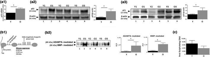
Rationale: Age-related changes in the intervertebral discs are the predominant contributors to back pain, a common physical and functional impairment experienced by older persons. Cellular senescence, a process wherein cells undergo growth arrest and chronically secrete numerous inflammatory molecules and proteases, has been reported to cause decline in the health and function of multiple tissues with age. Although senescent cells have been reported to increase in intervertebral degeneration (IDD), it is not known whether they are causative in age-related IDD.

Objective: The study aimed to elucidate whether a causal relationship exists between cellular senescence and age-related IDD.

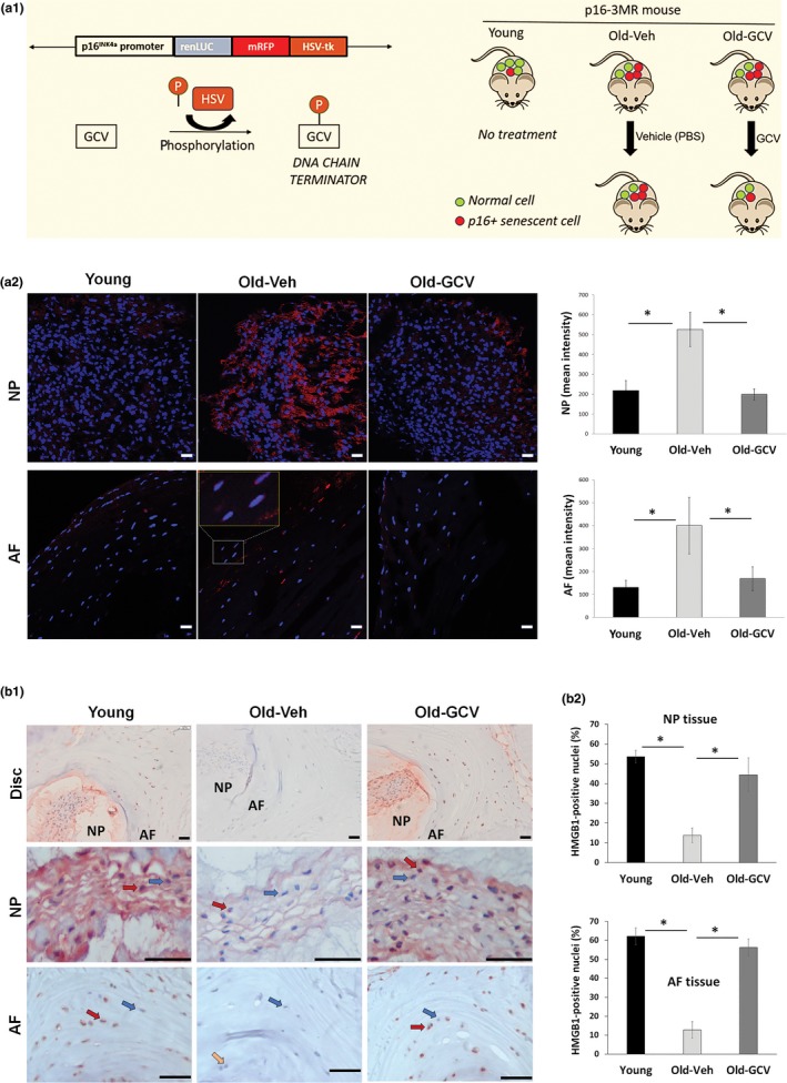
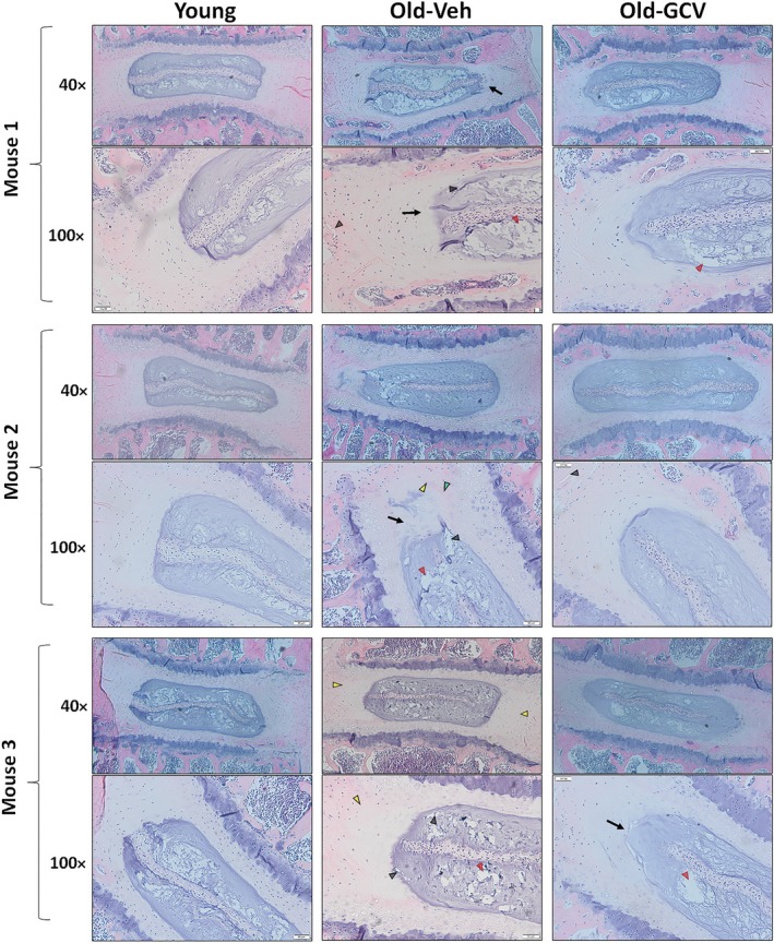
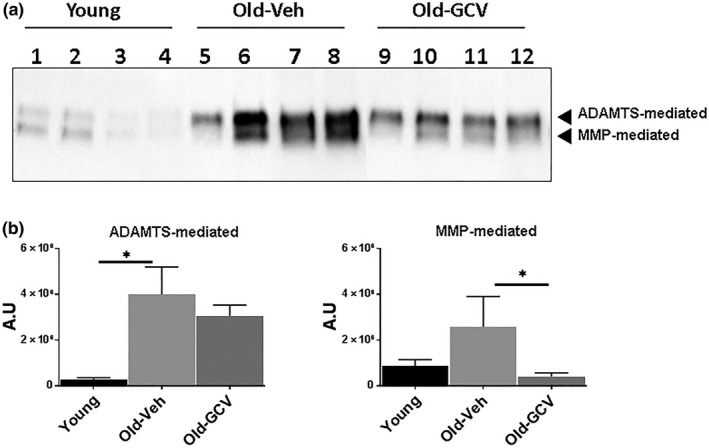
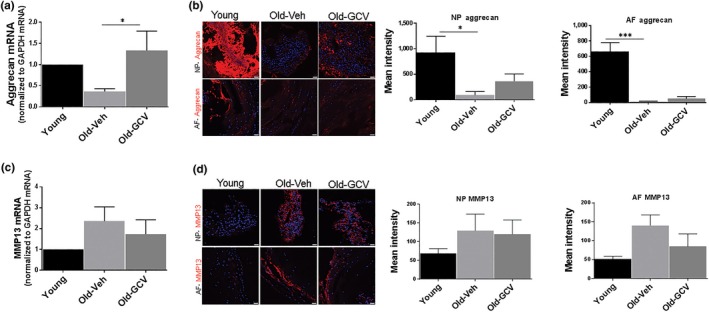
Methods and results: To examine the impact of senescent cells on age-associated IDD, we used p16-3MR transgenic mice, which enables the selective removal of p16Ink4a -positive senescent cells by the drug ganciclovir. Disc cellularity, aggrecan content and fragmentation alongside expression of inflammatory cytokine (IL-6) and matrix proteases (ADAMTS4 and MMP13) in discs of p16-3MR mice treated with GCV and untreated controls were assessed. In aged mice, reducing the per cent of senescent cells decreased disc aggrecan proteolytic degradation and increased overall proteoglycan matrix content along with improved histological disc features. Additionally, reduction of senescent cells lowered the levels of MMP13, which is purported to promote disc degenerative changes during aging.

Conclusions: The findings of this study suggest that systemic reduction in the number of senescent cells ameliorates multiple age-associated changes within the disc tissue. Cellular senescence could therefore serve as a therapeutic target to restore the health of disc tissue that deteriorates with age.

Keywords: aggrecanolysis; aging; cellular senescence; intervertebral disc; p16Ink4a; proteoglycan.


Conflict of interest statement

None declared.

Figures

Figure 1
Figure 1
Degenerative changes in intervertebral discs of naturally aged mice. (a) Expression of selected senescent markers, p16Ink4a (a1), p53 (a2) and p21 (a3) in disc tissue from young (6 month) and old (22 month) mice was determined by Western blotting and qRT–PCR. Graphs on the right of the Western images are quantification of results (a2 and a3) whereby the volume of respective protein band divided by volume of β‐actin band. (b) Increased disc aggrecan proteolysis with age. (b1) A schematic of the mouse aggrecan core protein covalently linked to the sulphate‐rich glycosaminoglycan (GAG) and noncovalently bound to a hyaluronic acid (HA) chain via the link protein. The cleavage sites between G1 and G2 interglobular domains by ADAMTS (G1‐NVTEGE392) and MMP (G1VDIPEN360) proteases are indicated. (b2) Western blot analysis of aggrecan fragments generated by ADAMTS and MMP proteases with graphs showing quantification results. Representative Western blots for three young and three old mice were shown (panels a2, a3, b2). (c) Proteoglycan synthesis as measured by 35S‐sulphate incorporation using whole disc organ cultures. Student's t test was used for parametric data (graphs a1, a2, b2), and the Mann–Whitney test was used for two‐variable nonparametric data (graphs a3, c). Data are means ± SD of four independent experiments (4 mice) for graphs a1, a3, c and three mice for graphs a2, b2. *p < 0.05. Y = young and O = old. Representative Western images of three different young (lanes 1, 3, 5) and old (lanes 2, 4, 6) mice are shown in panels a2, a3, and b2)
Figure 2
Figure 2
Clearance of senescent cells in p16‐3MR mice. (a1) Schematic of the p16‐3MR transgene and strategy used to selectively kill senescent cells. p16‐3MR mice were sacrificed at 12 months of age (Young) or were administered PBS (Old‐Veh) or GCV (Old‐GCV) for an additional 12 months before sacrifice. Confirmation of elimination of p16Ink4a‐positive cells in Old‐GCV mice was assessed by examining levels of RFP fluorescence (panel a2) and nuclear HMGB1 protein by IHC (panel b) in AF and NP tissues. Graphs on the right are quantification of the imaging results. Panel b2 shows quantitation of nuclear HMGB1 in disc cells. Data shown are means ± SEM of 4 independent experiments (4 mice), *p < 0.05. Examples of cells stained negative (blue arrows), partially positive (yellow arrow) or positive (red arrows) for nuclear HMGB1 are indicated. Scale bars = 10 µm (panel a2) and 50 µm (panel b1)
Figure 3
Figure 3
Impact of GCV treatment on gross morphology. H&E staining of lumbar disc was performed to assess the gross morphological changes with aging and GCV treatment. Compared to discs of young mice, discs of old p16‐3MR mice treated with PBS exhibited increasing loss of distinct NP/AF boundary (black arrows), loss of AF structure in which the AF lamellae become less concentric and more serpentine with each lamellae spaced farther apart (yellow arrowheads), loss of NP matrix indicated by large empty space gaps (red arrowheads) and fissures/clefts (black arrowheads). These degradative changes were blunted in the old p16‐3MR mice treated with GCV. Disc sections from three representative mice of each group (p16‐3MR Young, Old‐Veh and Old‐GCV) are shown. Scale bar = 50 μm of H&E‐stained disc sections
Figure 4
Figure 4
Effects of GCV treatment on disc aggrecanolysis of p16‐3MR mice. (a) Immunoblot of MMP‐ and ADAMTS‐mediated cleavage of aggrecan of discs of young mice (lanes 1–4), vehicle‐treated old mice (lanes 5–8) and GCV‐treated old mice (lanes 9–12). Graphs on right are quantification of aggrecan fragments shown in panel on left. Data shown are mean ± SD of 4 independent experiments, *p < 0.05
Figure 5
Figure 5
Effects of GCV treatment on aggrecan and MMP13 mRNA and protein levels in intervertebral discs of p16‐3MR mice. Aggrecan mRNA levels were quantified by qRT–PCR (a), and protein was quantified by immunofluorescence signals (b) in nucleus pulposus and annulus fibrosus section of disc tissue. MMP13 expression of whole disc mRNA by qRT–PCR (c) and protein by immunofluorescence (d) in inner nucleus pulposus and outer annulus fibrosus section of disc tissue. Graphs on the right are quantification of the immunofluorescence results. Data shown are mean ± SEM of four independent experiments (4 mice), *p < 0.05, ***p < 0.001. Scale bar = 10 μm

References

    1. Alvarez‐Garcia, O. , Matsuzaki, T. , Olmer, M. , Masuda, K. , & Lotz, M. K. (2017). Age‐related reduction in the expression of FOXO transcription factors and correlations with intervertebral disc degeneration. Journal of Orthopaedic Research: Official Publication of the Orthopaedic Research Society, 35, 2682–2691. - PMC - PubMed
    1. Baker, D. J. , Childs, B. G. , Durik, M. , Wijers, M. E. , Sieben, C. J. , Zhong, J. , & van Deursen, J. M. (2016). Naturally occurring p16(Ink4a)-positive cells shorten healthy lifespan. Nature, 530(7589), 184–189. 10.1038/nature16932 - DOI - PMC - PubMed
    1. Baker, D. J. , Wijshake, T. , Tchkonia, T. , LeBrasseur, N. , Childs, B. , van de Sluis, B. , … van Deursen, J. (2011). Clearance of p16Ink4a‐positive senescent cells delays ageing‐associated disorders. Nature, 479, 232–236. 10.1038/nature10600 - DOI - PMC - PubMed
    1. Buckwalter, J. A. (1995). Aging and degeneration of the human intervertebral disc. Spine, 20(11), 1307–1314. 10.1097/00007632-199506000-00022 - DOI - PubMed
    1. Campisi, J. (2005). Senescent cells, tumor suppression, and organismal aging: Good citizens, bad neighbors. Cell, 120(5), 513–522. 10.1016/j.cell.2005.02.003 - DOI - PubMed

Publication types

MeSH terms