Review
. 2019 Sep;235(3):590-615.
doi: 10.1111/joa.12948. Epub 2019 Mar 22.

Astrogliogenesis in human fetal brain: complex spatiotemporal immunoreactivity patterns of GFAP, S100, AQP4 and YKL-40

Affiliations
Review

Astrogliogenesis in human fetal brain: complex spatiotemporal immunoreactivity patterns of GFAP, S100, AQP4 and YKL-40

Camilla Bjørnbak Holst et al. J Anat. 2019 Sep.

Abstract

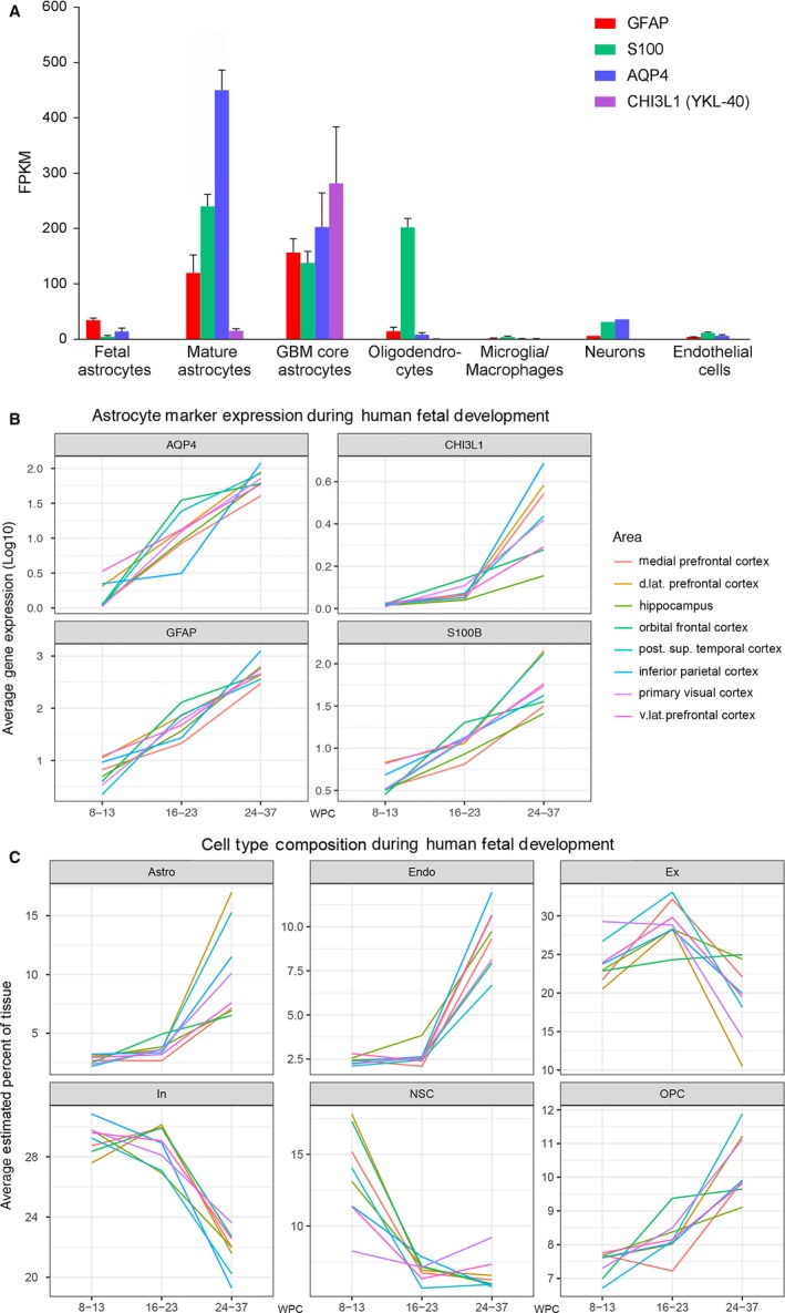
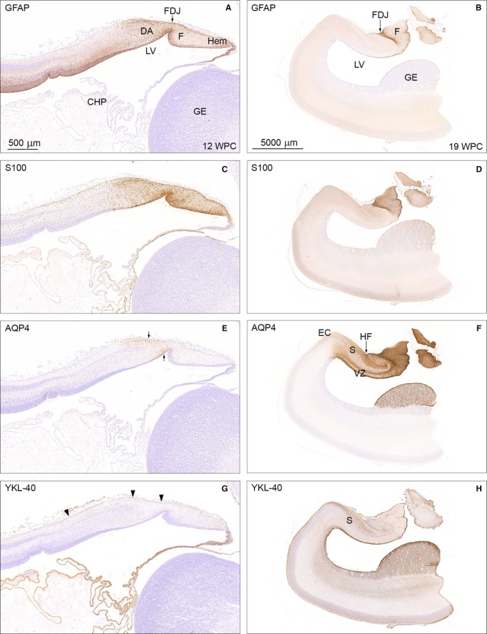
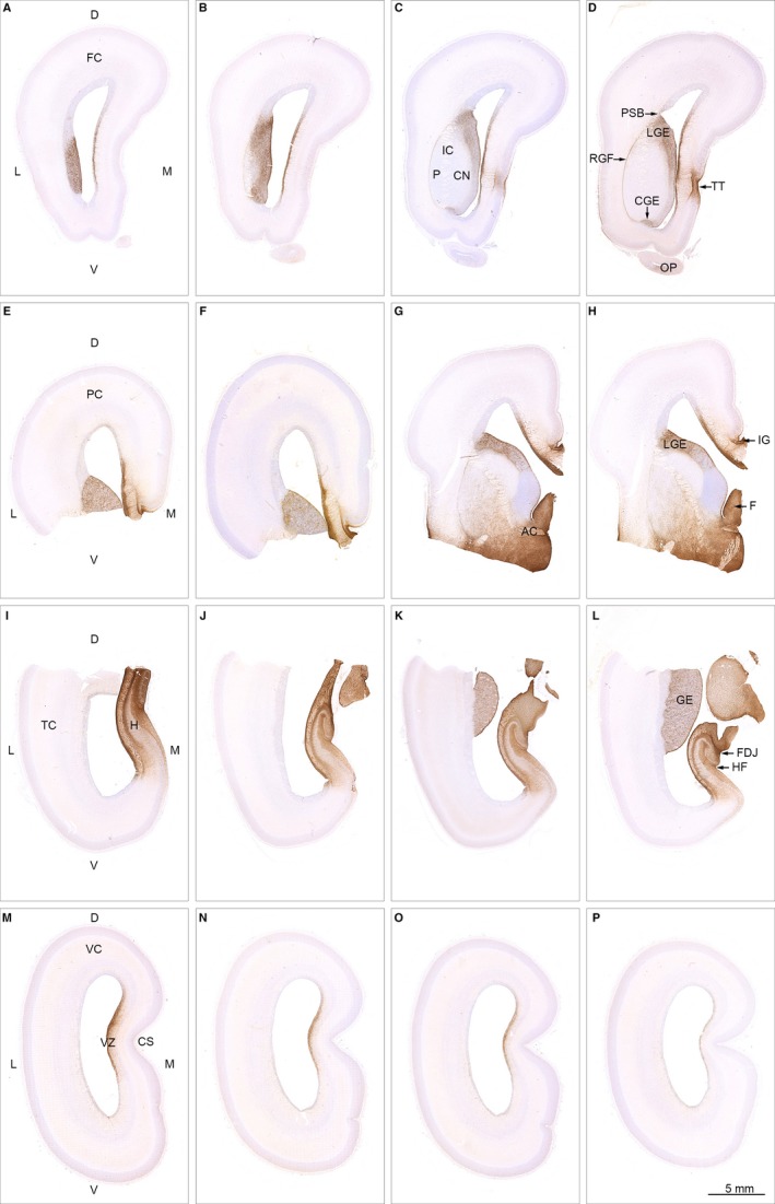
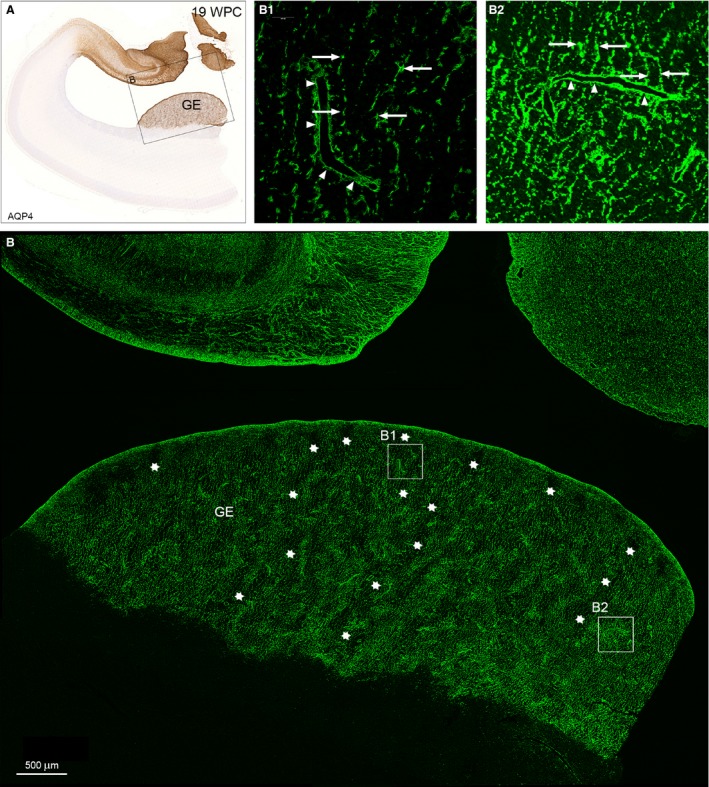
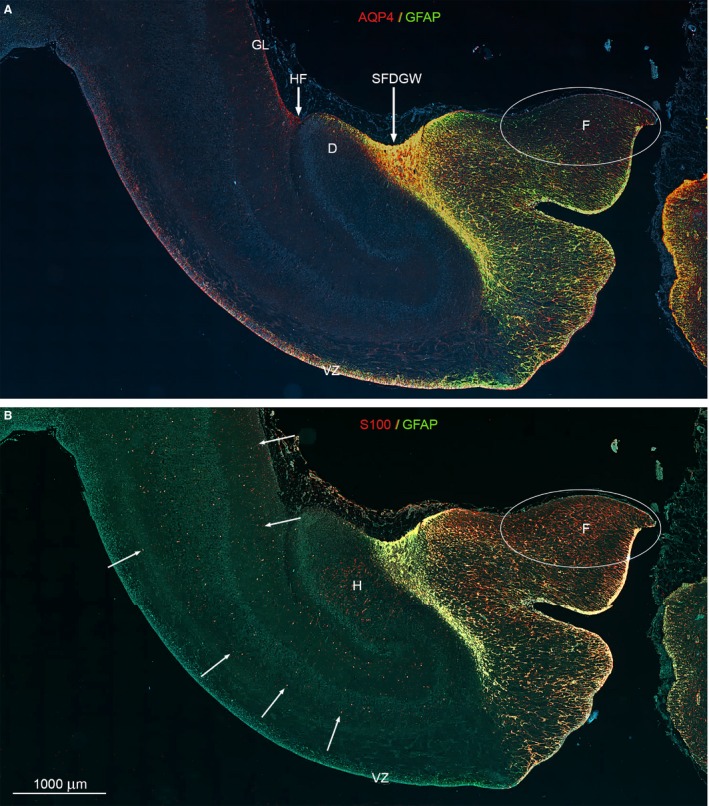
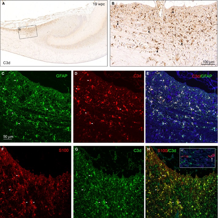
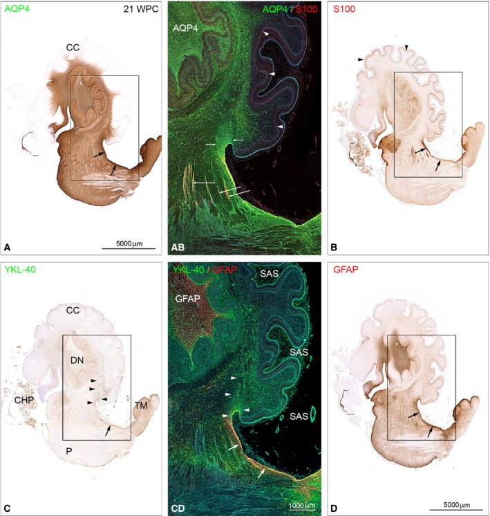
The astroglial lineage consists of heterogeneous cells instrumental for normal brain development, function and repair. Unfortunately, this heterogeneity complicates research in the field, which suffers from lack of truly specific and sensitive astroglial markers. Nevertheless, single astroglial markers are often used to describe astrocytes in different settings. We therefore investigated and compared spatiotemporal patterns of immunoreactivity in developing human brain from 12 to 21 weeks post conception and publicly available RNA expression data for four established and potential astroglial markers - GFAP, S100, AQP4 and YKL-40. In the hippocampal region, we also screened for C3, a complement component highly expressed in A1-reactive astrocytes. We found diverging partly overlapping patterns of the established astroglial markers GFAP, S100 and AQP4, confirming that none of these markers can fully describe and discriminate different developmental forms and subpopulations of astrocytes in human developing brain, although AQP4 seems to be the most sensitive and specific marker for the astroglial lineage at midgestation. AQP4 characterizes a brain-wide water transport system in cerebral cortex with regional differences in immunoreactivity at midgestation. AQP4 distinguishes a vast proportion of astrocytes and subpopulations of radial glial cells destined for the astroglial lineage, including astrocytes determined for the future glia limitans and apical truncated radial glial cells in ganglionic eminences, devoid of GFAP and S100. YKL-40 and C3d, previously found in reactive astrocytes, stain different subpopulations of astrocytes/astroglial progenitors in developing hippocampus at midgestation and may characterize specific subpopulations of 'developmental astrocytes'. Our results clearly reflect that lack of pan-astrocytic markers necessitates the consideration of time, region, context and aim when choosing appropriate astroglial markers.

Keywords: astrocytes; astrogliogenesis; glial markers; human brain development; radial glial cells.


Conflict of interest statement

The authors have no conflict of interest to declare.

Figures

Figure 1
Figure 1
RNA expression of GFAP, S100B,AQP4 and CHI3L140 in human developing and adult brain and estimation of cell‐type proportions in developing human brain. (A) RNA expression of GFAP, S100B,AQP4 and CHI3L1 (YKL‐40) in specific cell types in samples from human healthy temporal cortex (8–63 years, n = 12 for astrocytes, = 5 for oligodendrocytes, = 3 for microglia/macrophages, n = 1 for neurons, n = 2 for endothelial cells), astrocytes from fetal brain (16–16.5 wpc, n = 6) and astrocytes from glioblastoma tumor core (59–65 years, n = 3). Expression is listed as mean ± SEM (standard error of the mean) in FPKM. The RNA‐seq data in (A) was generously shared by the Barres Lab and is based on specific cell‐type suspensions derived by immunopanning (www.brainrnaseq.org) (Zhang et al. 2016). (B) is based a different dataset, consisting of RNA‐seq quantification of different brain regions in developing human brain and shows average gene expression (log10) (y‐axis) of GFAP, S100B, AQP4 and CHI3L1 (YKL‐40) (subplots) across all samples within three age intervals (early, mid‐ and late prenatal) (x‐axis) for selected brain regions (colors). Within the same dataset (C) shows average cell‐type proportions (y‐axis) for major cell types (subplots) within the same age groups (x‐axis) and regions (colors) as listed in (B). Specification of cell types (Box 2) is based on single‐cell RNA‐seq performed by Fan et al. (2018). RNA‐seq data for (B) and (C) originates from www.brainspan.org. For clarity, only cell types identified as making up more than 10% of cells, in at least one of the regions analyzed, are shown. Astro, astrocytes; d. lat, dorsolateral; Endo, endothelial cells; Ex, excitatory neurons; In, inhibitory neurons; NSC, neural stem cells; OPC, oligodendrocyte progenitor cells; post. sup, posterior superior; v.lat, ventrolateral.
Figure 2
Figure 2
Adjacent horizontal sections of forebrain including thalamus (T) and brainstem of a 12 wpc (CRL: 74 mm) human fetal brain stained for GFAP (A) and S100 (B) shown at low magnification. (A) Note the GFAP‐positive staining of the entire VZ except the GE. Also that CHP and midline raphe (R, arrow) are negative. The framed area is shown in higher magnification in Fig. 3A. (B) The neighboring section immunostained for S100 depicts absence of S100‐reactivity in VZ outside the immunopositive DA and fimbria. The midline raphe (R) of the brainstem shows strong immunoreactivity for S100. The boxed area is shown in higher magnification in Fig. 3C together with other astroglial markers. CHP, choroid plexus of the lateral ventricle; DA, dentate anlage; F, fimbria; GE, ganglionic eminence; LV, lateral ventricle; R, raphe; T, thalamus; VZ, ventricular zone. A and B – same magnification. Scale bar: (A) 5000 μm.
Figure 3
Figure 3
Temporal patterns of GFAP, S100, AQP4 and YKL‐40 immunoreactivity in 12 wpc (CRL: 74 mm) and 19 wpc (CRL: 165 mm) human fetal brain. Panel 1 (A,C,E,G) shows higher magnification of the framed areas in Figs 2A and B and of neighboring sections from the same 12 wpc fetus stained for AQP4 and YKL‐40. Panel 2 (B,D,F,H) illustrates the same region from a 19 wpc coronally sectioned fetus stained for the same astroglial markers: GFAP, S100, AQP4 and YKL‐40. At 12 wpc (panel 1) GFAP‐ and S100 immunoreactivity in (A) and (C) of the fimbria (F) and the dentate anlage (DA) separated by the fimbriodentate junction (FDJ) is strong but not entirely overlapping. In the marginal zone, S100‐reactivity continues into the hippocampus proper, which is devoid of GFAP staining. AQP4 and YKL‐40 immunoreactivity in (E) and (G) depicts the upcoming AQP4‐positive radial glial fibers in the DA between small arrows in (E) and the YKL‐40‐positive radial glial end feet (arrowheads) in (G). At 19 wpc the FDJ is strongly immunoreactive for GFAP (B), S100 (D) and AQP4 (F) but not for YKL‐40 (H). CHP, choroid plexus; DA, dentate anlage; EC, entorhinal cortex; F, fimbria; FDJ, fimbriodentate junction; GE, ganglionic eminence; Hem, hem; HF, hippocampal fissure; LV, lateral ventricle; S, subiculum; VZ, ventricular zone. Panel 1 (A, C, E and G) – same magnification, scale bar in (A): 500 μm. Panel 2 (B,D,F,H) – same magnification. Scale bar: (B) 5000 μm.
Figure 4
Figure 4
Spatial distribution of AQP4 in 19 wpc (CRL: 165 mm) human fetal brain. Sixteen sections immunostained for AQP4 from a series of more than 4000 coronal sections through the entire brain of a 19 wpc human fetus and taken from the material shown in Box 1 C (A–D from frontal lobe; E‐H from parietal lobe; I‐L from temporal lobe; M‐P from occipital lobe). At 19 wpc, APQ4 immunoreactivity delineates a continuous water transport system from the hippocampal formation (H) in (I) to taenia tecta (TT) in the medial frontal cortex (D) and via the basal forebrain (G,H) to the ganglionic eminence. The ventricular zone (VZ in M) of the medial wall also shows a continuous labeling from frontal to occipital lobe. The olfactory peduncle (OP in D) is weakly stained at this stage. Orientation: D, dorsal; L, lateral; M, medial; V, ventral. The entire cortical wall: FC, frontal cortex; PC, parietal cortex; TC, temporal cortex; VC, visual cortex. Subregions: AC, anterior commissure; CGE, caudal ganglionic eminence; CN, caudate nucleus; CS, calcarine sulcus; FDJ, fibriodentate junction; GE, ganglionic eminence; RGF, radial glia fiber fascicle; H, hippocampus; HF, hippocampal fissure; IC, internal capsule; IG, indusium griseum; LGE, lateral ganglionic eminence; OP, olfactory peduncle; PSB, pallial–subpallial boundary; TT, taenia tecta; VZ, ventricular zone. A‐P: same magnification. Scale bar: (P) 5 mm.
Figure 5
Figure 5
Distribution of AQP4 in the ganglionic eminence from a 19 wpc human fetal brain. A neighboring section to that of hippocampus and adjacent temporal cortex in Fig. 4K immunostained for AQP4 for bright‐field light microscopy (A) and confocal laser scanning microscopy (B). The framed area in (A) includes the ganglionic eminence (GE) in the lateral temporal wall and part of the strongly reacting hippocampal formation facing the lateral ventricle in the medial wall, which will be dealt with in Fig. 6. The GE in (B) is characterized by an uneven distribution of AQP4‐positive radial glial fibers resulting in a sponge‐like compartmentalized structure characterized by an intricate system resembling tunnels with thin and thick walls. The interior of some of the worm‐like tortuous tunnels is indicated by rows of white asterisks and shown in higher magnification in (B1). The more dense structure of the walls is depicted in (B2). (B1) Regularly spaced thin radial glial fibers with evenly spaced varicosities (arrows) characterize the tortuous tunnels of the AQP4‐positive ganglionic eminence. Side branches of the radial glial fibers ensheathe blood vessels (arrowheads) as astroglial end feet, although they are still part of the radial glial fiber system. (B2) The matrix of the sponge‐like structure possesses a more dense network of thick, rather coarse radial glial fibers with larger and more intensively stained varicosities (arrows), many of which seem to terminate on blood vessels as astroglial end feet (arrowheads). GE, ganglionic eminence. Scale bar: (B) 500 μm.
Figure 6
Figure 6
Whole‐slide fluorescent scanning of hippocampal formation from 19 wpc human fetal brain. Two immunofluorescent consecutive sections double‐labeled with antibodies against AQP4 and GFAP (A) and S100 and GFAP (B). Both sections are from the hippocampal formation adjacent to the bright‐field AQP4‐immunostained section shown in Fig. 5. (A, B) Include the entire wall of the hippocampal formation with the subpial fimbriodentate gliogenic wedge (SFDGW) region at the fimbriodentate junction and subiculum in the upper left corner – compare with Fig. 3F. The double‐labeled sections with DAPI‐stained nuclei shown in blue were subjected to whole‐slide fluorescent scanning. Radial glial cells in the ventricular zone (VZ) of the entire HF including the most distal part of fimbria is strongly positive for AQP4 as shown by the red and yellow color in (A). Within the cerebral wall, a diffuse fine dotted reactivity for AQP4 (red) can be distinguished in (A) (see also Fig. 3F) as opposed to the evenly distributed marked cellular S100 reactivity (red) indicated with white arrows in (B). The ventricular zone and the underlying future alveus are not labeled but there is an accumulation of S100‐positive cells in the hilus (H) of the dentate (D). (A, B) The subpial fimbriodentate gliogenic wedge (SFDGW) exhibits very strong immunoreactivity for the astroglial markers AQP4, GFAP and S100 and is mainly yellow with overlap, but also shows specific red (AQP4) patches in (A) and green (GFAP) patches in (B). D, dentate; F, fimbria; GL, glia limitans; H, hilus; HF, hippocampal fissure; SFDGW, subpial fimbriodentate gliogenic wedge; VZ, ventricular zone. (A–B) – same magnification. Scale bar: (B) 1000 μm.
Figure 7
Figure 7
C3d‐immunoreactive astrocytes of the subpial fimbriodentate gliogenic wedge. Bright‐field (A, B) and double‐immunofluorescence microscopy (C–H) of adjacent coronal sections of hippocampus from the subpial fimbriodentate gliogenic wedge described in Fig. 6 immunostained with antibodies against complement C3d (A, B), C3d and GFAP(C–E) and C3d and S100 (F–H). (A) Shows an accumulation of C3d‐immunoreactive cells in the subpial fimbriodentate gliogenic wedge. The boxed area shown in higher magnification in (B), demonstrates numerous C3d‐positive astrocytes (arrowheads). Double staining for GFAP (green) and C3d (red) in (C) and (D) shows a strong overlap of immunoreactive astrocytes in (E). Individual cells are labeled with 5 small white arrowheads. Double staining for S100 (red, F) and C3d (green, G) also shows a strong overlap of immunoreactive astrocytes (yellow) in (H). Four individual cells are labeled with small arrowheads demonstrating that early developing S100‐positive astrocytes possess C3d. The insert in (H) labeled (H’) depicts two more mature S100‐positive but C3d‐negative astrocytes (one indicated by small white arrow) from the middle of the neocortical wall. Scale bar in (B): 100 μm. (C‐H) Same magnification. Scale bar: (C) 50 μm.
Figure 8
Figure 8
Distribution of AQP4, S100 and YKL‐40, GFAP in brainstem and cerebellum from a 21 wpc (CRL: 200 mm) human fetal brain. Adjacent sagittal sections immunostained with antibodies against AQP4, S100 (A,B and AB) and YKL‐40, GFAP (C,D and CD). Note the marked differences in staining patterns with the entire brainstem and the core of cerebellum showing strong AQP4 immunoreactivity in contrast to a lack of staining of the cerebellar cortex (CC) in (A), whereas Bergman glia in cortex (arrowheads) in (B) exhibits a pronounced staining for S100. (C) A stream of YKL‐40‐positive cells migrates from the gliogenic hook between the dorsal brainstem and rostral cerebellum towards the center of cerebellum (arrowheads). All four astroglial markers show an overlapping pattern of immunoreactivity in the dorsal subpial gliogenic region (small black arrows) extending from the tectum mesencephali (TM in C) towards the cerebello‐mesencephalic junction. The boxed areas in (A‐D) are shown in (AB) and (CD) as double‐immunofluorescent sections stained with the same four antibodies and with nuclei labeled with DAPI shown in blue. The double‐labeled sections were subjected to whole‐slide fluorescent scanning and representative panels are displayed. (AB) and (CD) clearly demonstrate that GFAP‐, S100‐, AQP4‐ and YKL40‐positive cells represent separate although partly overlapping cell populations in the dorsal subpial gliogenic region (white arrows in CD). The radial glial cells of cerebellum (Bergmann glia) are strongly positively stained for S100 (arrowheads in AB and B) but not for the other astroglial markers. Cell aggregations seen along fiber tracts connecting cerebellum and upper brainstem seem to be distinctly immunoreactive for S100 (long white arrows in AB). The fiber tracts are surrounded by AQP4‐positive small cells (green). The migrating cell population from the dorsal subpial gliogenic region that is positively stained for YKL‐40 in (CD) (marked with white arrowheads) seems to be immunoreactive also for AQP4 (small white arrows in AB). Note the strongly GFAP‐immunoreactive core of the dentate nucleus (DN) in (CD) and a similar reactivity for AQP4 in AB. In the subarachnoid space (SAS) the numerous small leptomeningeal cells (green) and the smooth muscle cells (green) in small arteries are also YKL‐40‐positive. CC, cerebellar cortex; CHP, choroid plexus of the 4th ventricle; DN, dentate nucleus; P, pons; SAS, subarachnoid space; TM, tectum mesencephali. (A‐D) Same magnification. Scale bars: (A) 5000 μm; (AB,CD) same magnification; (CD) 1000 μm.

References

    1. Abbott NJ, Ronnback L, Hansson E (2006) Astrocyte‐endothelial interactions at the blood‐brain barrier. Nat Rev Neurosci 7, 41–53. - PubMed
    1. Abbott NJ, Pizzo ME, Preston JE, et al. (2018) The role of brain barriers in fluid movement in the CNS: is there a ‘glymphatic’ system? Acta Neuropathol 135, 387–407. - PubMed
    1. Abraham H, Perez‐Garcia CG, Meyer G (2004) p73 and Reelin in Cajal‐Retzius cells of the developing human hippocampal formation. Cereb Cortex 14, 484–495. - PubMed
    1. Abraham H, Vincze A, Jewgenow I, et al. (2010) Myelination in the human hippocampal formation from midgestation to adulthood. Int J Dev Neurosci 28, 401–410. - PubMed
    1. Allen NJ (2013) Role of glia in developmental synapse formation. Curr Opin Neurobiol 23, 1027–1033. - PubMed

Publication types

LinkOut - more resources