Review
. 2019 Jun 1;316(6):H1345-H1353.
doi: 10.1152/ajpheart.00096.2019. Epub 2019 Mar 22.

Role of T-cell activation in salt-sensitive hypertension

Affiliations
Review

Role of T-cell activation in salt-sensitive hypertension

Jiafa Ren et al. Am J Physiol Heart Circ Physiol. .

Abstract

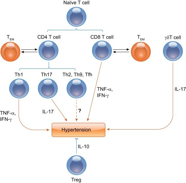
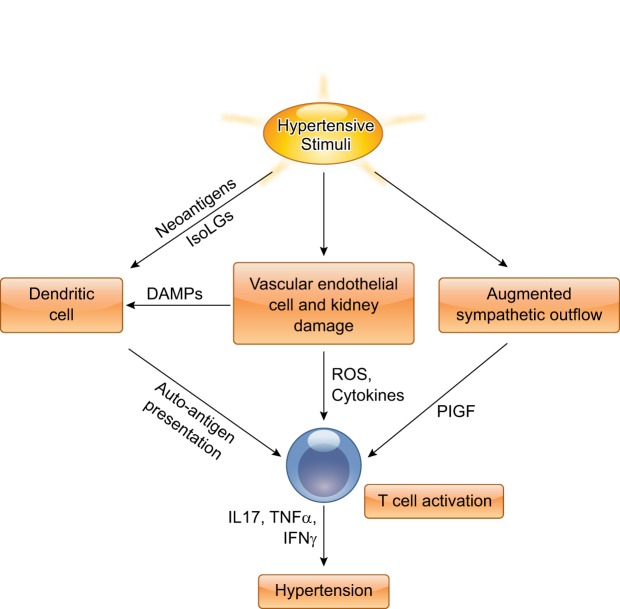
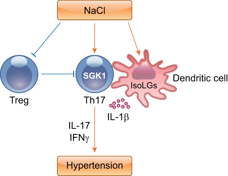
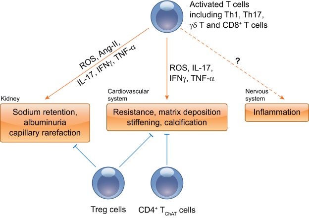
The contributions of T lymphocytes to the pathogenesis of salt-sensitive hypertension has been well established. Under hypertensive stimuli, naive T cells develop into different subsets, including Th1, Th2, Th17, Treg, and cytotoxic CD8+ T cells, depending on the surrounding microenviroment in organs. Distinct subsets of T cells may play totally different roles in tissue damage and hypertension. The underlying mechanisms by which hypertensive stimuli activate naive T cells involve many events and different organs, such as neoantigen presentation by dendritic cells, high salt concentration, and the milieu of oxidative stress in the kidney and vasculature. Infiltrating and activated T subsets in injured organs, in turn, exert considerable impacts on tissue dysfunction, including sodium retention in the kidney, vascular stiffness, and remodeling in the vasculature. Therefore, a thorough knowledge of T-cell actions in hypertension may provide novel insights into the development of new therapeutic strategies for patients with hypertension.

Keywords: T cell; end-organ damage; hypertension; salt.


Conflict of interest statement

No conflicts of interest, financial or otherwise, are declared by the authors.

Figures

Fig. 1.
Fig. 1.
Distinct T-lymphocyte subsets are crucial in hypertension. Th1, Th17, γδ T, and T regulatory (Treg) subpopulations promote or suppress the genesis of hypertension through proinflammatory cytokines, including TNF-α, IFN-γ, and IL-17, or anti-inflammatory cytokines such as IL-10, respectively. The relationships between Th2, Th9, or Tfh cells and hypertension are not clear and need to be further investigated. Some differentiated T-effector cells convert to memory T cells, and memory T cells will become T-effector cells again in antigen-specific recall responses.
Fig. 2.
Fig. 2.
Proposed mechanisms by which hypertensive stimuli activate T lymphocytes. Hypertensive stimuli, such as salt or ANG II, promote T-cell activation through various mechanisms. Oxidant stress induced by hypertensive stimuli causes the generation of neoantigens, such as isoketal adducts in dendritic cells. T cells recognize modified autoantigens presented by dendritic cells and then mature and differentiate into activated T cells, producing a variety of cytokines, including IL-17, TNF-α, and IFN-γ, eventually leading to the development of hypertension. Additionally, hypertensive stimuli cause vascular endothelial cell and kidney damage, and augmented sympathetic outflow, which can further activate T cells. ROS, reactive oxygen species; DAMPs, damage-associated molecular patterns; PIGF, placental growth factor.
Fig. 3.
Fig. 3.
High salt concentrations modulate T-cell differentiation. Increased sodium promotes the development of IL-17-producing CD4+ T cells in a DC-dependent or -independent manner. Moreover, high salt impairs immunosuppressive functions of Tregs. These events coordinately underpin hypertension.
Fig. 4.
Fig. 4.
Proposed roles of T cells in multiple organ dysfunction. Inflammatory-activated T cells infiltrate and accumulate in major target organs, including the kidney and vasculature, and then produce a variety of deleterious mediators to cause organ injury. The net effects of T-cell activation on sympathetic outflow in hypertension are complex and will require further study. Treg cells as anti-inflammatory cells normalize vascular and kidney function through anti-inflammatory cytokine IL-10. Another particular subset of CD4+ T cells, termed the CD4+ TChAT subset, plays a homeostatic role in vasorelaxation and regulation of blood pressure.

References

    1. Angel K, Provan SA, Gulseth HL, Mowinckel P, Kvien TK, Atar D. Tumor necrosis factor-alpha antagonists improve aortic stiffness in patients with inflammatory arthropathies: a controlled study. Hypertension 55: 333–338, 2010. doi:10.1161/HYPERTENSIONAHA.109.143982. - DOI - PubMed
    1. Banek CT, Knuepfer MM, Foss JD, Fiege JK, Asirvatham-Jeyaraj N, Van Helden D, Shimizu Y, Osborn JW. Resting afferent renal nerve discharge and renal inflammation: elucidating the role of afferent and efferent renal nerves in deoxycorticosterone acetate salt hypertension. Hypertension 68: 1415–1423, 2016. doi:10.1161/HYPERTENSIONAHA.116.07850. - DOI - PMC - PubMed
    1. Barbaro NR, Foss JD, Kryshtal DO, Tsyba N, Kumaresan S, Xiao L, Mernaugh RL, Itani HA, Loperena R, Chen W, Dikalov S, Titze JM, Knollmann BC, Harrison DG, Kirabo A. Dendritic cell amiloride-sensitive channels mediate sodium-induced inflammation and hypertension. Cell Reports 21: 1009–1020, 2017. doi:10.1016/j.celrep.2017.10.002. - DOI - PMC - PubMed
    1. Barhoumi T, Kasal DA, Li MW, Shbat L, Laurant P, Neves MF, Paradis P, Schiffrin EL. T regulatory lymphocytes prevent angiotensin II-induced hypertension and vascular injury. Hypertension 57: 469–476, 2011. doi:10.1161/HYPERTENSIONAHA.110.162941. - DOI - PubMed
    1. Caillon A, Mian MOR, Fraulob-Aquino JC, Huo KG, Barhoumi T, Ouerd S, Sinnaeve PR, Paradis P, Schiffrin EL. γδ T cells mediate angiotensin II-induced hypertension and vascular injury. Circulation 135: 2155–2162, 2017. doi:10.1161/CIRCULATIONAHA.116.027058. - DOI - PubMed

Publication types

MeSH terms

Substances