

Review
doi: 10.1016/j.sbi.2019.02.004.
Epub 2019 Mar 20.
AKTivation mechanisms
Affiliations
- PMID: 30901610
- PMCID: PMC6752985
- DOI: 10.1016/j.sbi.2019.02.004
Item in Clipboard
Review
AKTivation mechanisms
Curr Opin Struct Biol.
2019 Dec.
Abstract
Akt1-3 (Akt) are a subset of the AGC protein Ser/Thr kinase family and play important roles in cell growth, metabolic regulation, cancer, and other diseases. We describe some of the roles of Akt in cell signaling and the biochemical and structural mechanisms of the regulation of Akt catalysis by the phospholipid PIP3 and by phosphorylation. Recent findings highlight a diverse set of strategies to control Akt catalytic activity to ensure its normal biological functions.
Copyright © 2019 Elsevier Ltd. All rights reserved.
Figures
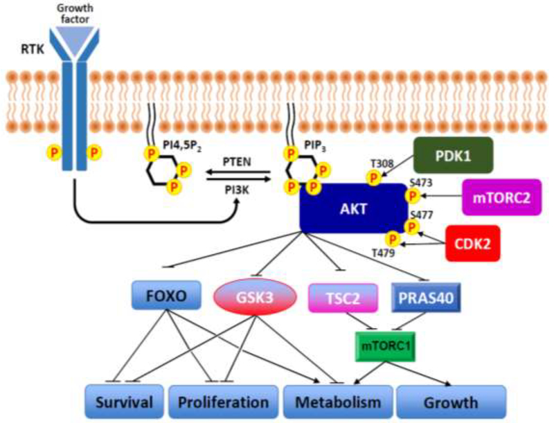
Upon activation by growth factors, PI3-K recruitment to a receptor tyrosine kinase leads to a transient increase in PIP3 levels. This phospholipid serves as an anchor point to recruit Akt1 to the plasma membrane where it undergoes phosphorylation at residues Thr308 and Ser473 by PDK1 and mTORC2, respectively. In addition, the more recently identified dual phosphorylations at residues Ser477 and Thr479 by Cdk2 that may occur in the cell nucleus can be an alternative mechanism for activating Akt1. Once activated, Akt1–3 phosphorylate downstream substrates that are involved in a diverse of cellular functions. Several of the well-characterized Akt1–3 substrates including FoxO, GSK3, TSC2 and PRAS40 are shown. P indicates phosphorylation.
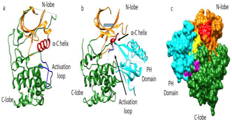
(a) Active conformation of Akt1 kinase domain aa144–480 (PDB: 3o96) where the N-lobe (orange), C-lobe (green), a-C helix (red) and activation loop (blue) are highlighted. (b) Inactive conformation of Akt31 aa1–443 (PDB: 6c0I) in complex with compound VIII (compound VIII not shown) highlighting the same features as in panel a. In addition, the PH domain (cyan) binds intramolecularly between the N and C lobes in the inactive state. (c) Surface representation of Akt1 in its inactive state (PDB: 6c0I) showing key residues in the interface of the PH and kinase domains. Potentially important residues mediating autoinhibition at the interface are highlighted: residues colored in yellow are part of the PH domain (E17, R23, N53, F55, L78, Q79, W80, T81) and residues in magenta are part of the kinase domain (V270, V271, L321, D323, D325, R328).
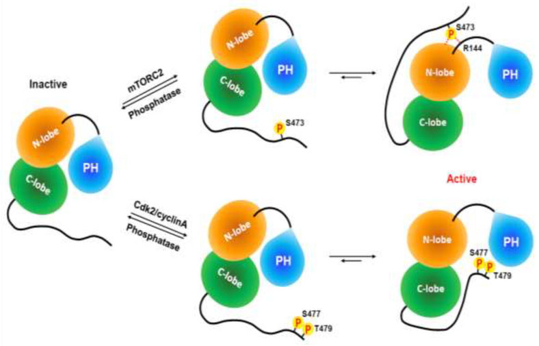
Without phosphorylation at its C-terminus, Akt1 has an autoinhibited conformation through a PH domain-kinase domain interaction. The interaction of phosphorylated Ser473 with the kinase domain N-lobe (Gln218) and the PH-kinase linker (Arg144) relieves tautoinhibition and activates Akt1. Dual phosphorylation of Ser477 and Thr479 also activates Akt1 by relieving PH domain-mediated autoinhibition via a different molecular mechanism.
References
-
- Parang K, Till JH, Ablooglu AJ, Kohanski RA, Hubbard SR, Cole PA: Mechanism-based design of a protein kinase inhibitor. Nat Struct Biol 2001, 8:37–41. - PubMed
Publication types
MeSH terms
Substances
Grants and funding
LinkOut - more resources
Full Text Sources
Miscellaneous
