Review
. 2019 Mar 21;11(3):288.
doi: 10.3390/v11030288.

Function, Architecture, and Biogenesis of Reovirus Replication Neoorganelles

Affiliations
Review

Function, Architecture, and Biogenesis of Reovirus Replication Neoorganelles

Raquel Tenorio et al. Viruses. .

Abstract

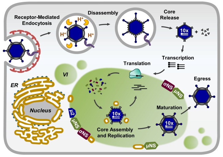
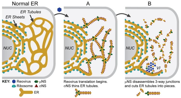
Most viruses that replicate in the cytoplasm of host cells form neoorganelles that serve as sites of viral genome replication and particle assembly. These highly specialized structures concentrate viral proteins and nucleic acids, prevent the activation of cell-intrinsic defenses, and coordinate the release of progeny particles. Reoviruses are common pathogens of mammals that have been linked to celiac disease and show promise for oncolytic applications. These viruses form nonenveloped, double-shelled virions that contain ten segments of double-stranded RNA. Replication organelles in reovirus-infected cells are nucleated by viral nonstructural proteins µNS and σNS. Both proteins partition the endoplasmic reticulum to form the matrix of these structures. The resultant membranous webs likely serve to anchor viral RNA⁻protein complexes for the replication of the reovirus genome and the assembly of progeny virions. Ongoing studies of reovirus replication organelles will advance our knowledge about the strategies used by viruses to commandeer host biosynthetic pathways and may expose new targets for therapeutic intervention against diverse families of pathogenic viruses.

Keywords: Reovirus; bluetongue virus; double-stranded RNA; endoplasmic reticulum; rotavirus; viral factories; viral inclusions; viral nonstructural proteins; viral replication organelles.


Conflict of interest statement

The authors declare no conflict of interest.

Figures

Figure 1
Figure 1
The reovirus replication cycle. VI—viral inclusions; ER—endoplasmic reticulum.
Figure 2
Figure 2
Reovirus inclusions imaged by light and electron microscopy. (AC) Human brain microvascular endothelial cells were infected with reovirus strain T1L M1-P208S for 24 h, and were fixed, permeabilized, and processed for immunofluorescence staining with a chicken anti-μNS polyclonal serum and a secondary antibody conjugated with Alexa 594 (green). This strain forms large, globular VIs. (A) The phase-contrast microscopy shows dark, dense, globular structures (asterisks) in the cytosol of reovirus-infected cells. (B) The localization of μNS by fluorescence microscopy confirms that the dense structures seen by phase-contrast microscopy are viral inclusions (asterisks). (C) The merging of phase-contrast and fluorescence microscopy images. (D,E) HeLa cells were infected with T1L M1-P208S and fixed at 24 h. (D) Ultrathin sections (~70 nm) of infected cells were imaged by transmission electron microscopy (TEM). A characteristic viral inclusion (VI) is shown. The VI contains mature virions (black arrowheads) and empty viral particles (white arrowheads). Mitochondria (mi), endoplasmic reticulum (ER) cisternae, and microtubules (arrows) surround the VI. N—nucleus. (E) VI as visualized by TEM of serial sections, 3D reconstruction, and image processing. The mitochondria (red) and ER cisternae (gold) surround a network of smooth membranes (light yellow) with mature virions (black) and empty viral particles (white). The nucleus is colored in blue and the microtubules in green. Scale bars are 10 μM in (AC) and 250 nm in (D,E).
Figure 3
Figure 3
Reovirus inclusions contain ER membranes. (A,B) HeLa cells were infected with reovirus T1L M1-P208S for 14 h, frozen in liquid nitrogen, and sectioned at −120 °C. The thawed cryosections were processed for immunogold labeling with primary antibodies specific for two ER proteins—protein disulfide isomerase (PDI) (A) and calreticulin (CLT) (B)—and for secondary antibodies conjugated with 10 nm colloidal gold particles. The rough ER (RER) cisternae around the VIs and membranes inside the VIs are labeled with antibodies specific for ER proteins (white arrows in A and B). These membranes are in close contact with the viral particles (black arrows in B). V—viral particle. (C) Electron tomography (ET) of a single VI. A thawed cryosection was processed by single-tilt-axis ET, 3D reconstruction, and image processing. The 3D model shows that the VI is a collection of vesicles and tubules with viral particles attached to membranes (white arrows). RER—yellow; viral particles—light blue; mitochondria—red; nuclear membrane—dark blue; tubules and membrane fragments inside the inclusion—brown; vesicles inside the inclusion—orange. Scale bars are 500 nm in (A) and 200 nm in (B,C). Modified from Tenorio et al., 2018 [31].
Figure 4
Figure 4
Model of ER remodeling and VI biogenesis. The ER in uninfected cells is composed of sheets and tubules. (A) In reovirus-infected cells, σNS binds to the ER tubules and transforms them into thin structures. (B) µNS binds to these thin tubules and triggers their fragmentation. Small tubules and vesicles coalesce to form the VI. The schematics at the bottom demonstrate how σNS and µNS might remodel the ER. NUC—nucleus. Modified from Tenorio et al., 2018 [31].

References

    1. Miller S., Krijnse-Locker J. Modification of intracellular membrane structures for virus replication. Nat. Rev. Microbiol. 2008;6:363–374. doi: 10.1038/nrmicro1890. - DOI - PMC - PubMed
    1. Novoa R.R., Calderita G., Arranz R., Fontana J., Granzow H., Risco C. Virus factories: Associations of cell organelles for viral replication and morphogenesis. Biol. Cell. 2005;97:147–172. doi: 10.1042/BC20040058. - DOI - PMC - PubMed
    1. Risco C., Fernandez de Castro I. Virus morphogenesis in the cell: Methods and observations. Subcell. Biochem. 2013;68:417–440. - PMC - PubMed
    1. Paul D., Bartenschlager R. Architecture and biogenesis of plus-strand RNA virus replication factories. World J. Virol. 2013;2:32–48. doi: 10.5501/wjv.v2.i2.32. - DOI - PMC - PubMed
    1. de Castro I.F., Volonte L., Risco C. Virus factories: Biogenesis and structural design. Cell. Microbiol. 2013;15:24–34. doi: 10.1111/cmi.12029. - DOI - PMC - PubMed

Publication types