. 2019 Mar 22;19(1):261.
doi: 10.1186/s12885-019-5437-3.

Molecular iodine exerts antineoplastic effects by diminishing proliferation and invasive potential and activating the immune response in mammary cancer xenografts

Affiliations

Molecular iodine exerts antineoplastic effects by diminishing proliferation and invasive potential and activating the immune response in mammary cancer xenografts

Irasema Mendieta et al. BMC Cancer. .

Abstract

Background: The immune system is a crucial component in cancer progression or regression. Molecular iodine (I2) exerts significant antineoplastic effects, acting as a differentiation inductor and immune modulator, but its effects in antitumor immune response are not elucidated.

Methods: The present work analyzed the effect of I2 in human breast cancer cell lines with low (MCF-7) and high (MDA-MB231) metastatic potential under both in vitro (cell proliferation and invasion assay) and in vivo (xenografts of athymic nude mice) conditions.

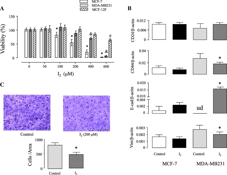
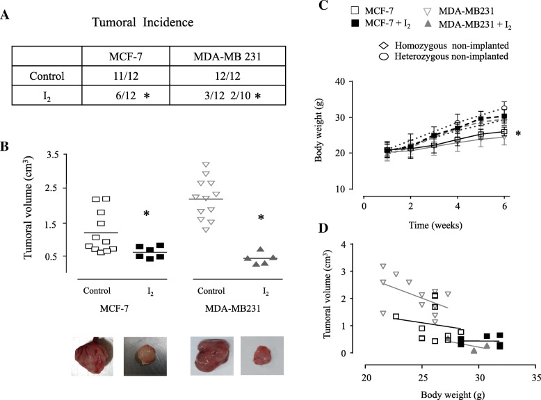
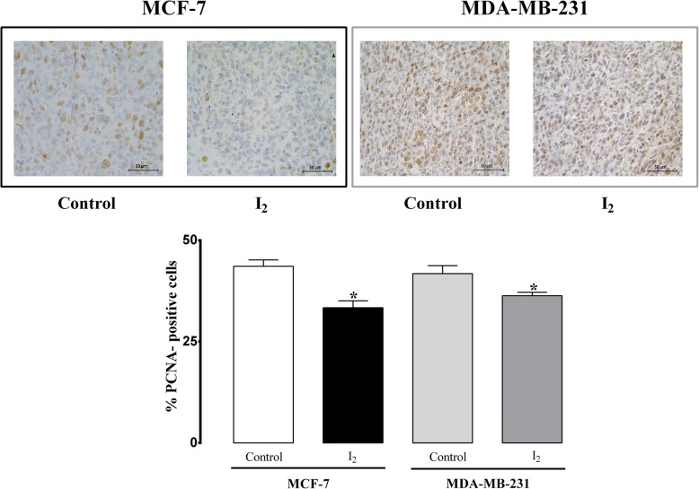
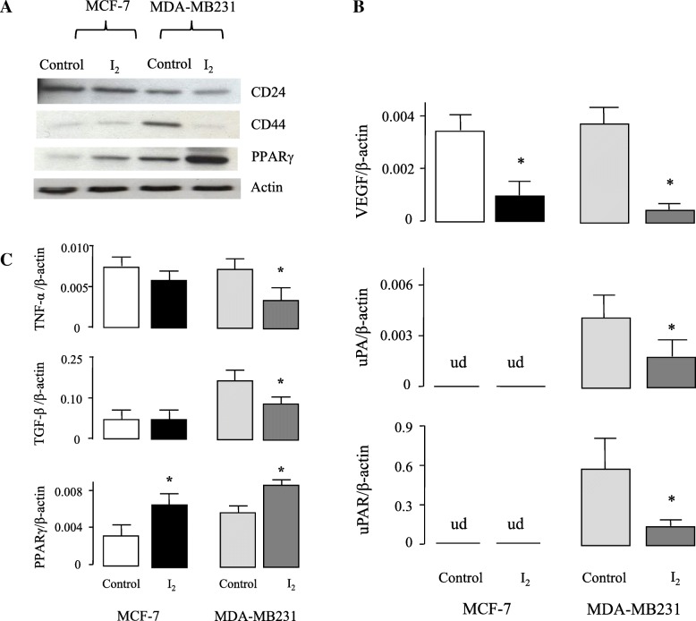
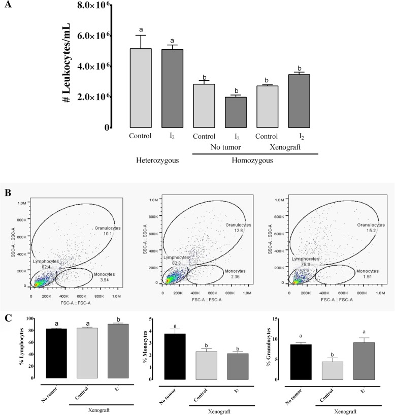
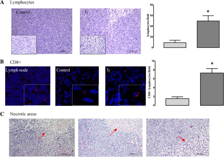
Results: In vitro analysis showed that the 200 μM I2 supplement decreases the proliferation rate in both cell lines and diminishes the epithelial-mesenchymal transition (EMT) profile and the invasive capacity in MDA-MB231. In immunosuppressed mice, the I2 supplement impairs implantation (incidence), tumoral growth, and proliferation of both types of cells. Xenografts of the animals treated with I2 decrease the expression of invasion markers like CD44, vimentin, urokinase plasminogen activator and its receptor, and vascular endothelial growth factor; and increase peroxisome proliferator-activated receptor gamma. Moreover, in mice with xenografts, the I2 supplement increases the circulating level of leukocytes and the number of intratumoral infiltrating lymphocytes, some of them activated as CD8+, suggesting the activation of antitumor immune responses.

Conclusions: I2 decreases the invasive potential of a triple negative basal cancer cell line, and under in vivo conditions the oral supplement of this halogen activates the antitumor immune response, preventing progression of xenografts from laminal and basal mammary cancer cells. These effects allow us to propose iodine supplementation as a possible adjuvant in breast cancer therapy.

Keywords: Immune response; Iodine; MCF-12F; MCF-7; MDA-MB231; Mammary cancer; Molecular iodine; PPARγ; Xenografts.


Conflict of interest statement

Ethics approval

All procedures followed the Animal Care and Use program (NIH, USA) and were approved by the Research Ethics Committee at INB (Protocol #035).

Competing interests

The authors declare that they have no competing interests.

Publisher’s Note

Springer Nature remains neutral with regard to jurisdictional claims in published maps and institutional affiliations.

Figures

Fig. 1
Fig. 1
Effect of molecular iodine (I2) on viability, epithelial-mesenchymal transition (EMT) and invasive potential in normal (MCF-12F) and cancerous (MCF-7 and MDA-MB231) mammary cells. a, Percentage of viable cells after incubation for 48 h in media containing iodine at the indicated concentrations, as determined by the 3-(4, 5-dimethylthiazol-2-yl)-2, 5-diphenyltetrazolium bromide (MTT) assay. Significant differences in * for mammary cancer MCF-7 cells, & for mammary cancer MDA-MB231 cells and in # for normal mammary epithelium MCF-12F cells (P < 0.05) between their respective control (one-way ANOVA and Tukey’s test). b, Cancerous cells incubated for 48 h in media containing 200 (MCF-7) and 400 (MDA-MB231) μM I2 were analyzed for mRNA expression for EMT markers CD24, CD44, E-cadherin (E-cad), and vimentin (Vim) by quantitative real-time PCR (RT-qPCR). Values were normalized to the amount of β-actin mRNA amplified. Ud, undetectable; *, significant differences vs. the control (Student’s t-test; P < 0.05). c, The invasiveness assay was performed to calculate the invasive capability of MDA-MB231 cells in presence of 200 μM I2. Images were taken at 20X magnification. Scale bar, 100 μm. Number of cells ± SD that penetrated the membrane. *Significant difference between treated and untreated MDA-MB231 cells (Student’s t-test, P < 0.05) (n = 3 individual samples)
Fig. 2
Fig. 2
Effect of I2 supplement on tumoral incidence, tumoral volume and body weight gain in animals with xenografts of cancer cells. Female athymic homozygous (Foxn1 nu/nu) mice were inoculated with 5 × 106 cells of each cell line in 50 μl PBS and 50 μl Matrigel. The drinking water and the water used for 0.025% I2 solution were always deionized. The water supplement (alone or with I2) began 48 h after cell inoculation and was maintained throughout the study. Parameters were analyzed after 6 weeks of inoculations. a, Tumoral incidence. Number of animals that presented observable tumorous mass (0.2 cm3). A second group of animals with 10 animals was incorporated to obtain five samples of xenografts from the MDA-MB231+ I2 group. * Significant differences between I2-treated mice and control mice using Chi-square test. b, Final tumoral volume. Each dot represents an individual tumor by each group. Data are expressed ± SD. * Significant difference (Student’s t-test; P < 0.05) between their respective control. Photographs of representative tumor mass for each group. c, Body weight gain. Lines represent the body weight gain in animals implanted with xenografts and supplemented or not with 0.025% I2 for 6 weeks. The graph also shows the weight recorded for homo and heterozygous non-implanted animals (with and without I2 supplement) at 0, 3 and 6 weeks. Data are expressed ± SD. * Significant difference, one-way ANOVA and Tukey’s test. d, Correlation analysis. Linear regression between final tumor volume (cm3) and final body weight (6th week) in implanted homozygous mice with and without I2 supplement. P value for MCF-7, 0.6661; MCF-7 + I2, 0.9629; MDA-MB231, 0.0852; MDA-MB231 + I2, 0.2865. No significant differences were found
Fig. 3
Fig. 3
Proliferation rate. Immunohistochemistry showing the presence of PCNA-positive cells in xenografts from animals supplemented with or without 0.025% I2 in drinking water for six weeks. Percentage of PCNA-positive cells was quantified by counting the number of labeled cells in at least 500 cells per region, and three random regions were analyzed. Images were taken at 20X magnification. Data are expressed as the mean ± SD (n = 6). *Significantly different from its control (Student’s t-test; P < 0.05)
Fig. 4
Fig. 4
Effect of I2 supplement on expression of invasive and inflammatory markers in xenografts. Nude mice were supplemented with or without 0.025%I2 in drinking water for six weeks. a, Representative Western blot of CD24, CD44 and PPARγ proteins from MCF-7 and MDA-MB231 samples. Equal amounts of protein were loaded on each lane (50 μg), and Actin was run as a control for loading and exposure time (two independent experiments were performed). b and c, mRNA expression for vascular endothelial growth factor (VEGF), urokinase plasminogen activator (uPA), uPA receptor (uPAR), tumor necrosis factor alpha (TNFα), transforming growth factor beta (TGF-β), and peroxisome proliferator-activated receptor gamma (PPARγ) were analyzed by RT-qPCR. Values were normalized to the amount of β-actin mRNA amplified (n = 5–6 individual samples). Ud, undetectable. * Significant differences vs. the control (Student’s t-test; P < 0.05)
Fig. 5
Fig. 5
Effect of I2 supplement on circulating immune leucocytes. Six-week-old hetero- and homozygous nude mice (with and without MDA-MB231 xenografts) were supplemented with or without 0.025% I2 (in drinking water for 6 weeks. a, The number of leukocytes in peripheral blood was determined by direct counting after dilution with Turk’s solution. b, Peripheral blood populations of leukocytes were separated and quantified by FACS (cytometric images). Differential quantification (lower panel). Data are expressed as the mean ± SD (n = 8–10 animals per group). Different letters denote statistical differences (one-way ANOVA, Tukey’s test; P < 0.05)
Fig. 6
Fig. 6
Effect of I2 supplement on tumoral immune response. a, Micrographs stained with H&E (20X), insert 2X+ zoom of photograph shown lymphocytes (hyperpigmented cells). b, Confocal immunofluorescence (63X) of lymph node (positive control) and MDA-MB231 xenografts (Control and I2) with antibody against CD8 protein (red). Insert: zoom 2X+ to visualize CD8+ lymphocytes (red stain in cytoplasm). Nuclei were stained with DAPI (blue). The analysis was performed as the average of three random fields and the quantification was performed using the ImageJ 1.47 software. Data are expressed as the mean ± SD (n = 6). *Significantly different from control (Student’s t-test; P < 0.05). c, Representative micrographs from necrotic areas (red arrows) from MDA-MD231 + I2 xenografts (H&E, 20X)

Similar articles

Cited by

References

    1. Ferlay J, Soerjomataram I, Dikshit R, Eser S, Mathers C, Rebelo M, et al. Cancer incidence and mortality worldwide: sources, methods and major patterns in GLOBOCAN 2012. Int J Cancer. 2015;136(5):E359–EE86. doi: 10.1002/ijc.29210. - DOI - PubMed
    1. Charafe-Jauffret E, Ginestier C, Monville F, Finetti P, Adelaide J, Cervera N, et al. Gene expression profiling of breast cell lines identifies potential new basal markers. Oncogene. 2006;25(15):2273–2284. doi: 10.1038/sj.onc.1209254. - DOI - PubMed
    1. Duffy MJ, McGowan PM, Harbeck N, Thomssen C, Schmitt M. uPA and PAI-1 as biomarkers in breast cancer: validated for clinical use in level-of-evidence-1 studies. Breast Cancer Res. 2014;16(4):428. doi: 10.1186/s13058-014-0428-4. - DOI - PMC - PubMed
    1. Abdel-Rahman O. Targeting vascular endothelial growth factor (VEGF) pathway in iodine-refractory differentiated thyroid carcinoma (DTC): from bench to bedside. Crit Rev Oncol Hematol. 2015;94(1):45–54. doi: 10.1016/j.critrevonc.2014.11.009. - DOI - PubMed
    1. Kessenbrock K, Plaks V, Werb Z. Matrix metalloproteinases: regulators of the tumor microenvironment. Cell. 2010;141(1):52–67. doi: 10.1016/j.cell.2010.03.015. - DOI - PMC - PubMed

MeSH terms