

Molecular changes to tendons after collagenase-induced acute tendon injury in a senescence-accelerated mouse model
- PMID: 30902076
- PMCID: PMC6429773
- DOI: 10.1186/s12891-019-2488-1
Molecular changes to tendons after collagenase-induced acute tendon injury in a senescence-accelerated mouse model
Abstract
Background: Aging impairs tendon healing and is a potential risk factor for chronic tendinitis. During normal aging, tendons undergo structural and biomechanical degenerative changes, accompanied by a reduction in the number of tenocytes and changes to their properties. However, molecular changes in aged tendons under inflammatory conditions are not well understood. The present study analyzed the molecular changes in collagenase induced acute tendon injury using a senescence-accelerated mouse (SAM) model.
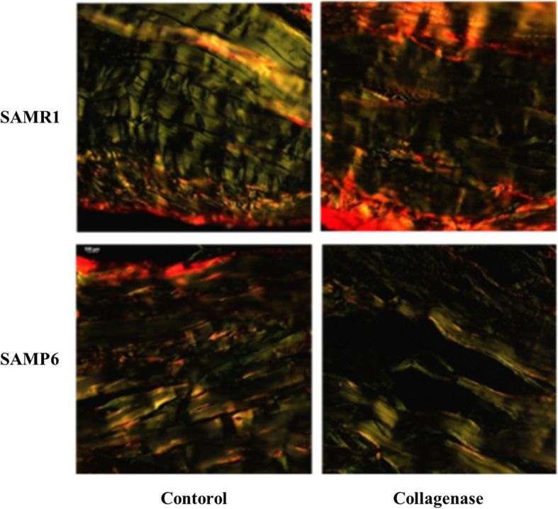
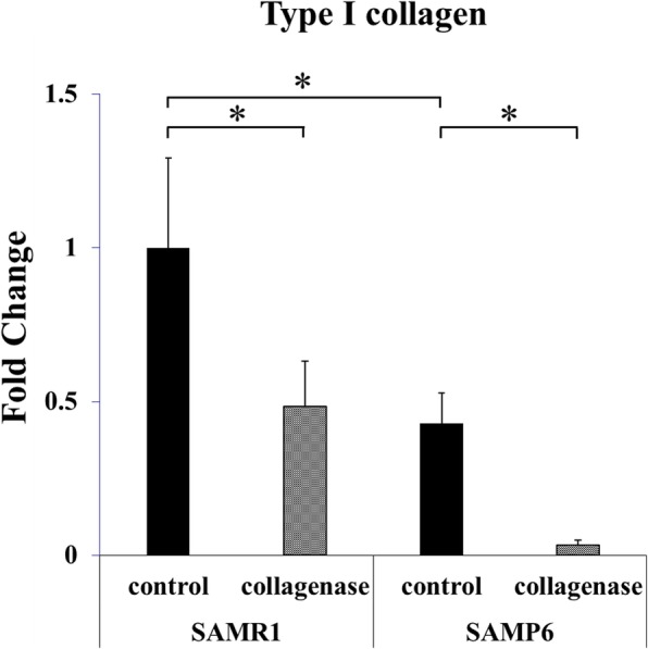
Methods: SAMP6 mice were used as an aging animal model and SAMR1 mice were used as a control to represent a senescence-resistant inbred strain. All the mice used in the study were 40 weeks old. Collagenase I from Clostridium histolyticum (20 μL) was injected percutaneously to the tendon-bone junction of the Achilles tendon. Two weeks after treatment, the Achilles tendons were harvested and stained using Picrosirius Red to determine collagen expression. Real-time PCR was performed to analyze gene expression of IL-6, tenomodulin, type I and type II collagen, MMP-9, TIMP-1, and TIMP-2.
Results: Collagenase injection resulted in significantly higher gene expression of IL-6 but significantly lower tenomodulin expression compared with the control in SAMP6 and SAMR1 mice. In SAMP6 mice, gene expression of type III collagen and MMP-9 was significantly higher in the collagenase-injected group compared with the control group. SAMP6 mice also showed lower expression of type I collagen, TIMP-1, and TIMP-2 in the collagenase-injected group compared with the control group. Picrosirius Red staining showed the highest expression of type III collagen in the collagenase-injected SAMP6 group compared with the other groups.
Conclusions: The collagenase-injected SAMP6 group showed higher expression of IL-6, MMP-9, and type III collagen and lower expression of type I collagen, TIMP-1, and TIMP-2, which are known to suppress metalloproteinases. The results indicate that aging may lead to dysfunction of the tendon healing process after acute tendon injury.
Keywords: Acute tendon injury; Aging; Tendon degeneration.
Conflict of interest statement
Ethics approval and consent to participate
Our IRB (Institutional Animal Care and Use Committee At kusunoki and Myodani Campus Kobe University) provided the approval for our study and the approval informations are follows. (Permission Number: P140610-R1). All animal procedures were performed under the approval and guidance of our IRB.
Consent for publication
Not applicable.
Competing interests
The authors declare that they have no competing interests.
Publisher’s Note
Springer Nature remains neutral with regard to jurisdictional claims in published maps and institutional affiliations.
Figures

Similar articles
-
Morphological changes of skeletal muscle, tendon and periosteum in the senescence-accelerated mouse (SAMP6): a murine model for senile osteoporosis.Tissue Cell. 2006 Oct;38(5):325-35. doi: 10.1016/j.tice.2006.08.001. Epub 2006 Sep 28. Tissue Cell. 2006. PMID: 17010403
-
Cytokine-induced tendinitis: a preliminary study in rabbits.J Orthop Res. 1999 Mar;17(2):168-77. doi: 10.1002/jor.1100170204. J Orthop Res. 1999. PMID: 10221832
-
High-fat diet-induced obesity exacerbated collagenase-induced tendon injury with upregulation of interleukin-1beta and matrix metalloproteinase-1.Connect Tissue Res. 2024 Nov;65(6):447-457. doi: 10.1080/03008207.2024.2409751. Epub 2024 Oct 4. Connect Tissue Res. 2024. PMID: 39364694
-
Characterization of senescence-accelerated mouse prone 6 (SAMP6) as an animal model for brain research.Exp Anim. 2014;63(1):1-9. doi: 10.1538/expanim.63.1. Exp Anim. 2014. PMID: 24521858 Free PMC article. Review.
-
Tendon - function-related structure, simple healing process and mysterious ageing.Folia Morphol (Warsz). 2018;77(3):416-427. doi: 10.5603/FM.a2018.0006. Epub 2018 Jan 18. Folia Morphol (Warsz). 2018. PMID: 29345715 Review.
Cited by
-
Models of tendon development and injury.BMC Biomed Eng. 2019;1:32. doi: 10.1186/s42490-019-0029-5. Epub 2019 Nov 29. BMC Biomed Eng. 2019. PMID: 32095779 Free PMC article.
-
Establishment of a Mouse Degenerative Model of Patellar Tendinopathy with Upregulation of Inflammation.Int J Mol Sci. 2024 Mar 29;25(7):3847. doi: 10.3390/ijms25073847. Int J Mol Sci. 2024. PMID: 38612656 Free PMC article.
-
Interleukin-6 upregulates extracellular matrix gene expression and transforming growth factor β1 activity of tendon progenitor cells.BMC Musculoskelet Disord. 2023 Nov 22;24(1):907. doi: 10.1186/s12891-023-07047-9. BMC Musculoskelet Disord. 2023. PMID: 37993850 Free PMC article.
-
Tendon Extracellular Matrix Assembly, Maintenance and Dysregulation Throughout Life.Adv Exp Med Biol. 2021;1348:45-103. doi: 10.1007/978-3-030-80614-9_3. Adv Exp Med Biol. 2021. PMID: 34807415 Review.
-
A Novel Tendon Injury Model, Induced by Collagenase Administration Combined with a Thermo-Responsive Hydrogel in Rats, Reproduces the Pathogenesis of Human Degenerative Tendinopathy.Int J Mol Sci. 2024 Feb 3;25(3):1868. doi: 10.3390/ijms25031868. Int J Mol Sci. 2024. PMID: 38339145 Free PMC article.
References
MeSH terms
Substances
LinkOut - more resources
Full Text Sources
Medical
Research Materials
Miscellaneous
