. 2019 Mar 22;18(1):33.
doi: 10.1186/s12938-019-0654-1.

Multiscale experimental study on the effects of different weight-bearing levels during moderate treadmill exercise on bone quality in growing female rats

Affiliations

Multiscale experimental study on the effects of different weight-bearing levels during moderate treadmill exercise on bone quality in growing female rats

Juan Fang et al. Biomed Eng Online. .

Abstract

Background: Bone tissue displays a hierarchical organization. Mechanical environments influence bone mass and structure. This study aimed to explore the effects of different mechanical stimuli on growing bone properties at macro-micro-nano scales.

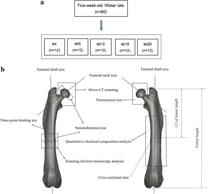
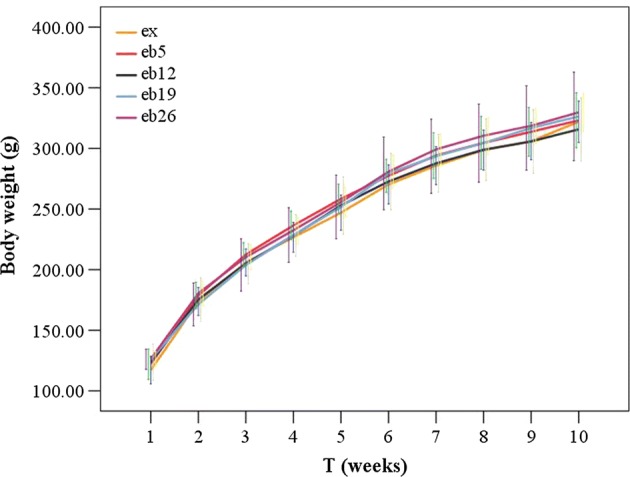
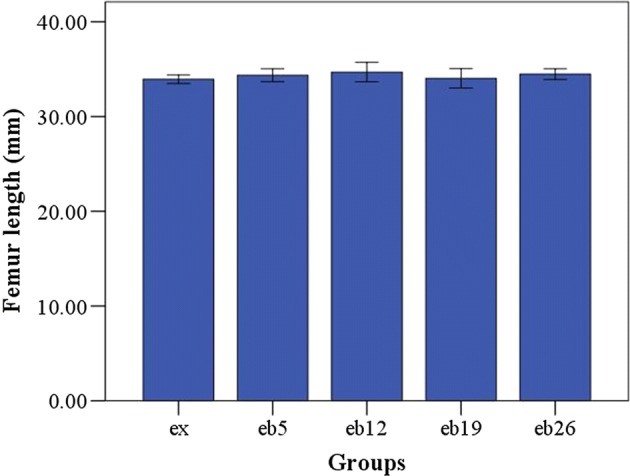
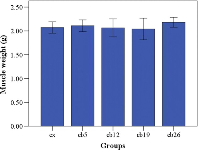
Methods: Sixty five-week-old female Wistar rats were treadmill exercised at moderate intensity with the speed of 12 m/min, and then randomly divided into five groups according to weight-bearing level. After 8 weeks of experiment, femurs were harvested to perform multiscale tests.

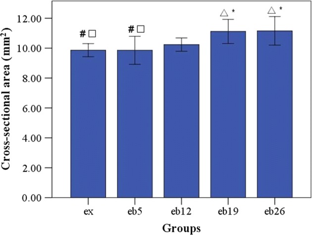
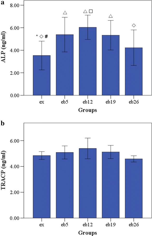
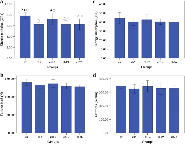
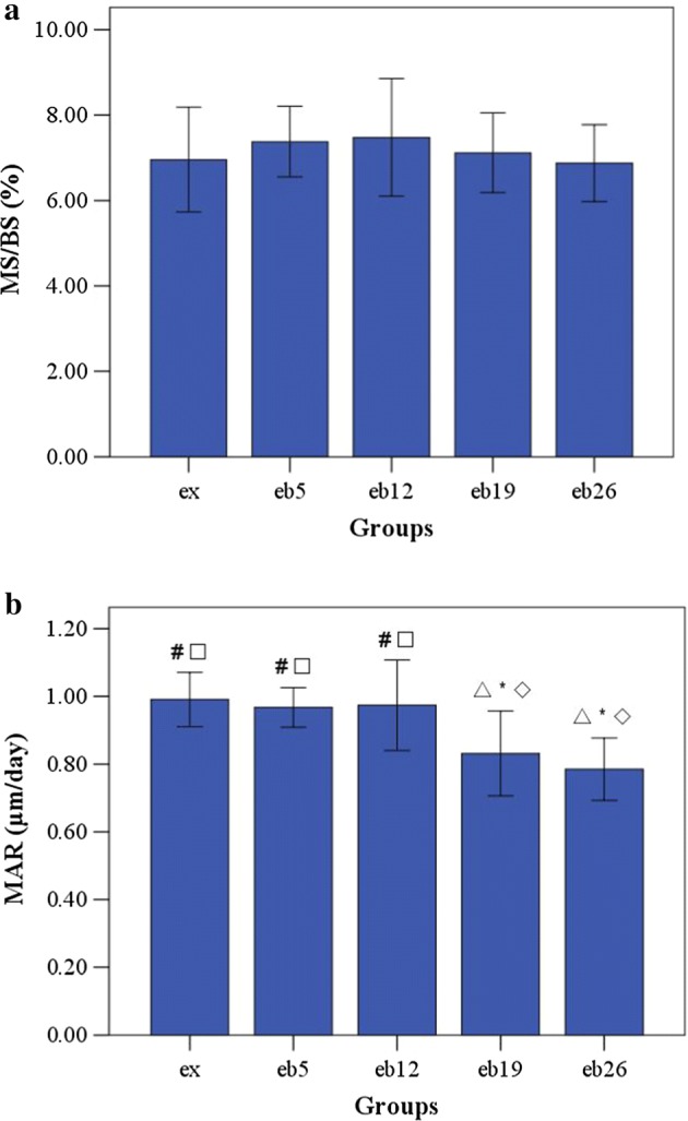
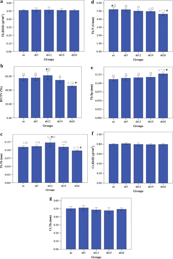
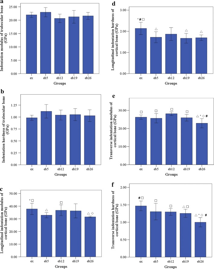
Results: Bone formation was significantly increased by weight-bearing exercise, whereas bone resorption was not significantly inhibited. Trabecular and cortical bone mineral densities showed no significant increase by weight-bearing exercise. The microstructure of trabecular bone was significantly improved by 12% weight-bearing exercise. However, similar positive effects were not observed with further increase in weight-bearing levels. The nanomechanical properties of trabecular bone were not significantly changed by weight-bearing exercise. The macrostrength of whole femur and the nanomechanical properties of cortical bone significantly decreased in the 19% and 26% weight-bearing exercise groups.

Conclusion: When rats ran on the treadmill at moderate intensity during growth period, additional 12% weight-bearing level could significantly increase bone formation, improve microstructure of trabecular bone, as well as maintain the structure and mechanical properties of cortical bone. Excessive weight-bearing level caused no positive effects on the trabecular bone microstructure and properties of cortical bone at all scales. In addition, increased weight-bearing level exerted no significant influence on trabecular and cortical bone mineral densities.

Keywords: Bone quality; Moderate intensity; Treadmill exercise; Weight-bearing.


Conflict of interest statement

The authors declare that they have no competing interests.

Figures

Fig. 1
Fig. 1
The schematic of experimental grouping and the measurements performed in the femur. a Experimental grouping. b Measurement of the femur
Fig. 2
Fig. 2
Indentation sites for nanoindentation test. Actual indented sites were marked by red cross under optical microscopy, and the sample thickness was 2 mm
Fig. 3
Fig. 3
The body weight of each group during the experiment. Error bars represent standard deviation (SD). At T2, the 8-week exercise program started
Fig. 4
Fig. 4
The cross-sectional areas in the middle segment of femoral shaft at the end of experiment. Error bars represent SD. Statistically different from the ex group (p < 0.05); *Statistically different from the eb5 group (p < 0.05); Statistically different from the eb12 group (p < 0.05); #Statistically different from the eb19 group (p < 0.05); Statistically different from the eb26 group (p < 0.05)
Fig. 5
Fig. 5
The length of femur. Error bars represent SD
Fig. 6
Fig. 6
The calf muscles weight. Error bars represent SD
Fig. 7
Fig. 7
The serum concentrations of ALP and TRACP in each group. Error bars represent SD. Statistically different from the ex group (p < 0.05); *Statistically different from the eb5 group (p < 0.05); Statistically different from the eb12 group (p < 0.05); #Statistically different from the eb19 group (p < 0.05); Statistically different from the eb26 group (p < 0.05). a ALP concentration. b TRACP concentration
Fig. 8
Fig. 8
The macromechanical parameters of the left femur measured by three-point bending mechanical test. Error bars represent SD. Statistically different from the ex group (p < 0.05); *Statistically different from the eb5 group (p < 0.05); Statistically different from the eb12 group (p < 0.05); #Statistically different from the eb19 group (p < 0.05); Statistically different from the eb26 group (p < 0.05). a Elastic modulus. b Failure load. c Energy absorption. d Stiffness
Fig. 9
Fig. 9
The MS/BS and MAR of trabecular bone in the femoral head. Error bars represent SD. MAR mineral apposition rate, MS/BS the ratio of mineralizing surface to bone surface. Statistically different from the ex group (p < 0.05); *Statistically different from the eb5 group (p < 0.05); Statistically different from the eb12 group (p < 0.05); #Statistically different from the eb19 group (p < 0.05); Statistically different from the eb26 group (p < 0.05). a MS/BS, b MAR
Fig. 10
Fig. 10
The 3D microstructure parameters of trabecular bone in femoral head and cortical bone in femoral shaft. Error bars represent SD. Tb.BMD trabecular bone mineral density, BV/TV bone volume fraction, Tb.Th trabecular thickness, Tb.N trabecular number, Tb.Sp trabecular separation, Ct.BMD cortical bone mineral density, Ct.Th cortical bone thickness. Statistically different from the ex group (p < 0.05); *Statistically different from the eb5 group (p < 0.05); Statistically different from the eb12 group (p < 0.05); #Statistically different from the eb19 group (p < 0.05); Statistically different from the eb26 group (p < 0.05). a Tb.BMD, b BV/TV, c Tb.Th, d Tb.N, e Tb.Sp, f Ct.BMD, g Ct.Th
Fig. 11
Fig. 11
The indentation modulus and hardness of trabecular and cortical bone sites as determined by nanoindentation. Error bars represent SD. ETb.L longitudinal indentation modulus of trabecular bone, HTb.L longitudinal hardness of trabecular bone, ECt.L longitudinal indentation modulus of cortical bone, HCt.L longitudinal hardness of cortical bone, ECt.T transverse indentation modulus of cortical bone, HCt.T transverse hardness of cortical bone. Statistically different from the ex group (p < 0.05); *Statistically different from the eb5 group (p < 0.05); Statistically different from the eb12 group (p < 0.05); #Statistically different from the eb19 group (p < 0.05); Statistically different from the eb26 group (p < 0.05). a ETb.L of trabecular bone, b HTb.L of trabecular bone, c ECt.L of cortical bone, d HCt.L of cortical bone, e ECt.T of cortical bone, f HCt.T of cortical bone

References

    1. Rico H, Gervas JJ, Hernandez R, Seco C, Villa FL, Revilla M, et al. Effects of alprazolam supplementation on vertebral and femoral bone mass in rats on strenuous treadmill training exercise. Calcif Tissue Int. 1999;65:139–142. doi: 10.1007/s002239900672. - DOI - PubMed
    1. Yeh JK, Aloia JF, Barilla ML. Effects of 17β-estradiol replacement and treadmill exercise on vertebral and femoral bones of the ovariectomized rat. Bone Miner. 1994;24:223–234. doi: 10.1016/S0169-6009(08)80139-0. - DOI - PubMed
    1. Yuan Y, Chen X, Zhang LL, Wu JN, Guo JM, Zou DC, et al. The roles of exercise in bone remodeling and in prevention and treatment of osteoporosis. Prog Biophys Mol Biol. 2016;122:122–130. doi: 10.1016/j.pbiomolbio.2015.11.005. - DOI - PubMed
    1. Ju YI, Sone T, Ohnaru K, Choi HJ, Fukunaga M. Differential effects of jump versus running exercise on trabecular architecture during remobilization after suspension-induced osteopenia in growing rats. J Appl Physiol. 2012;112:766–772. doi: 10.1152/japplphysiol.01219.2011. - DOI - PubMed
    1. Warner SE, Shea JE, Miller SC, Shaw JM. Adaptations in cortical and trabecular bone in response to mechanical loading with and without weight bearing. Calcif Tissue Int. 2006;79:395–403. doi: 10.1007/s00223-005-0293-3. - DOI - PubMed

LinkOut - more resources