. 2019 Apr 10;25(4):602-616.e7.
doi: 10.1016/j.chom.2019.02.013. Epub 2019 Mar 19.

NLRP12 Regulates Anti-viral RIG-I Activation via Interaction with TRIM25

Affiliations

NLRP12 Regulates Anti-viral RIG-I Activation via Interaction with TRIM25

Szu-Ting Chen et al. Cell Host Microbe. .

Abstract

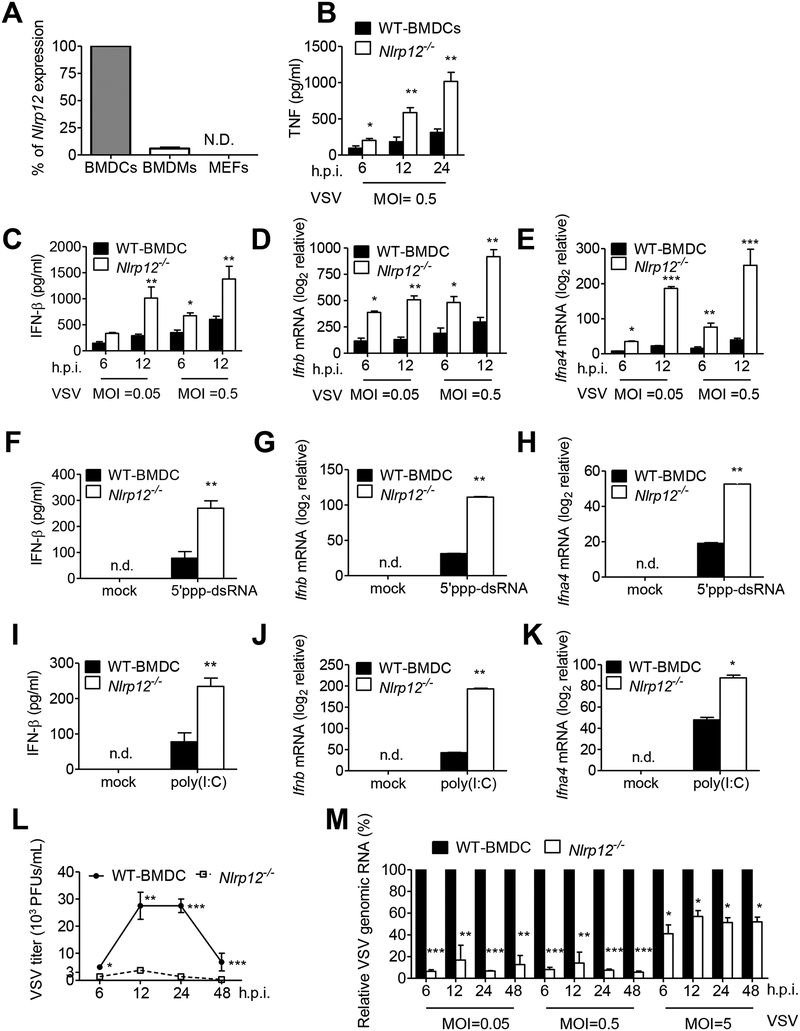
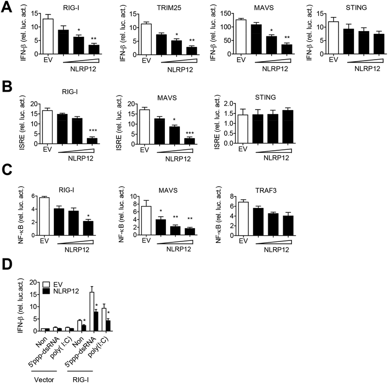
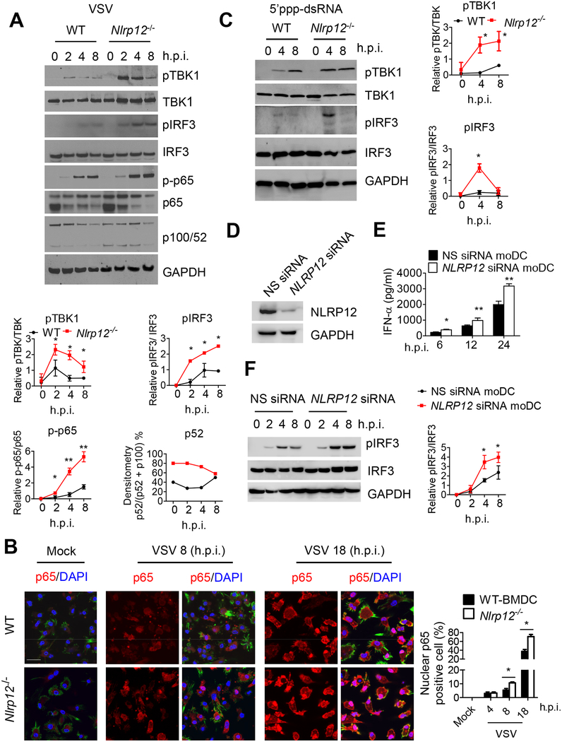
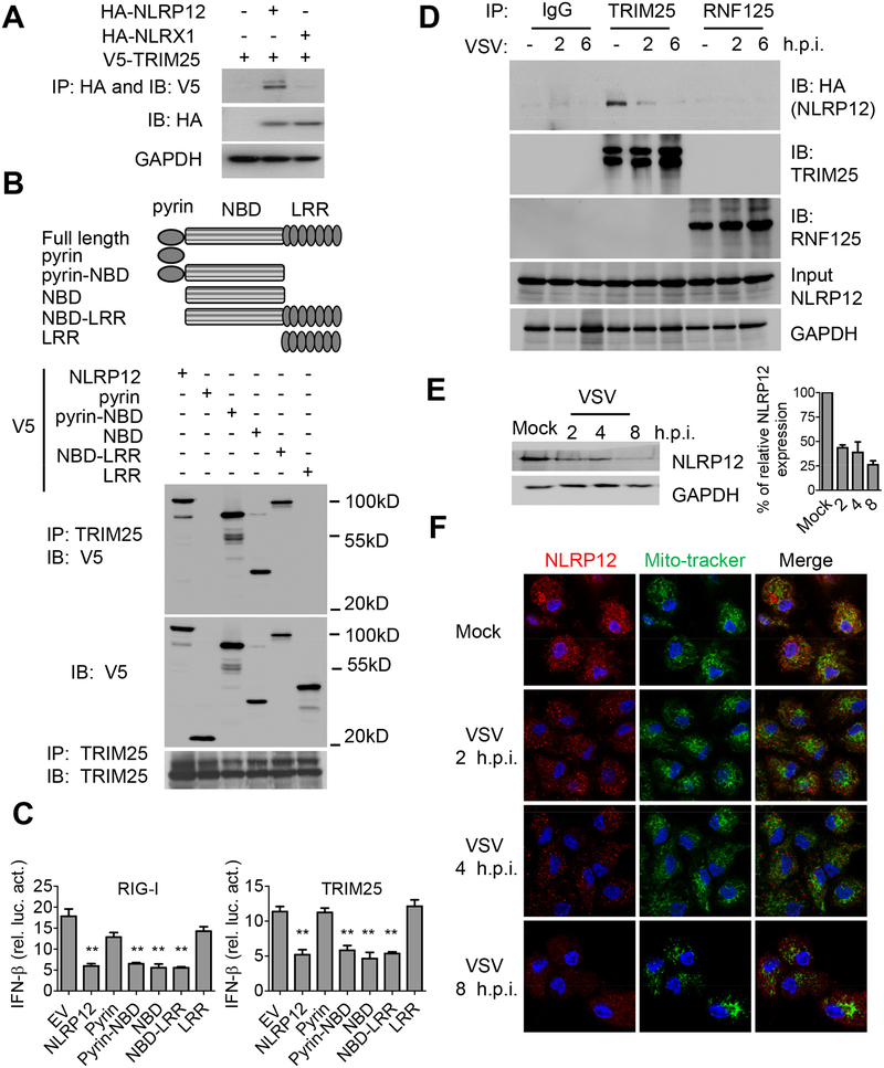
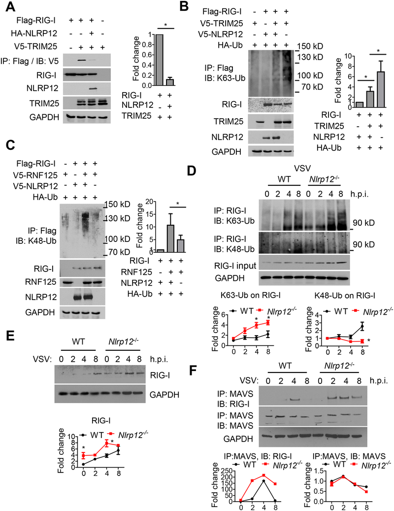
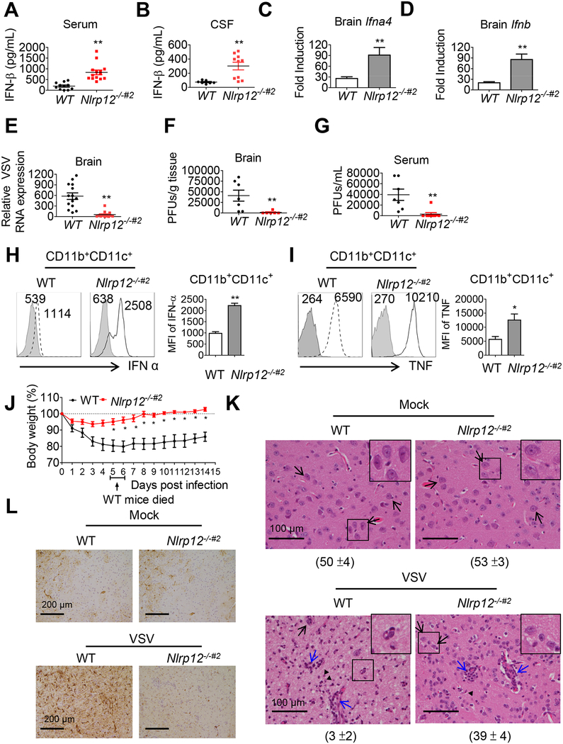
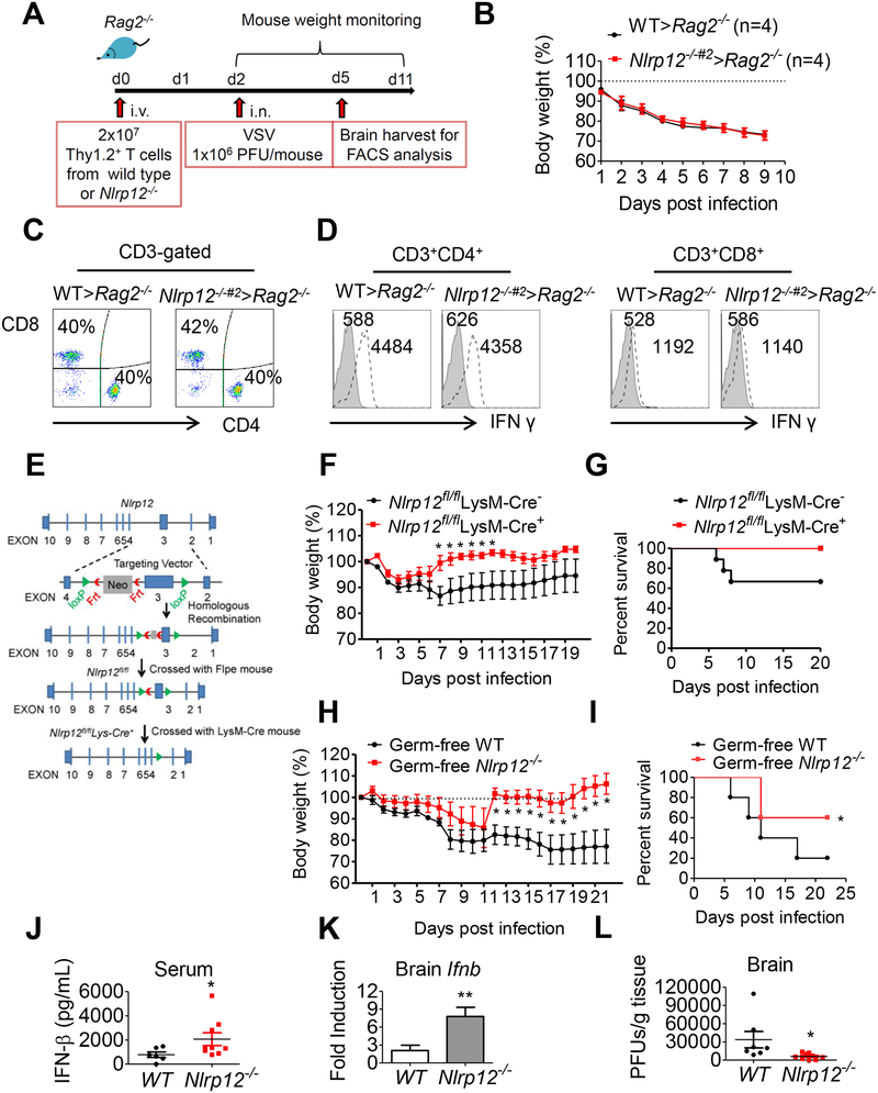
Establishing the balance between positive and negative innate immune mechanisms is crucial for maintaining homeostasis. Here we uncover the regulatory crosstalk between two previously unlinked innate immune receptor families: RIG-I, an anti-viral cytosolic receptor activated type I interferon production, and NLR (nucleotide-binding domain, leucine repeat domain-containing protein). We show that NLRP12 dampens RIG-I-mediated immune signaling against RNA viruses by controlling RIG-I's association with its adaptor MAVS. The nucleotide-binding domain of NLRP12 interacts with the ubiquitin ligase TRIM25 to prevent TRIM25-mediated, Lys63-linked ubiquitination and activation of RIG-I. NLRP12 also enhances RNF125-mediated, Lys48-linked degradative ubiquitination of RIG-I. Vesicular stomatitis virus (VSV) infection downregulates NLRP12 expression to allow RIG-I activation. Myeloid-cell-specific Nlrp12-deficient mice display a heightened interferon and TNF response and are more resistant to VSV infection. These results indicate that NLRP12 functions as a checkpoint for anti-viral RIG-I activation.


Conflict of interest statement

COMPETING FINANCIAL INTERESTS

The authors declare no competing financial interests.

Figures

Figure 1.
Figure 1.. NLRP12 suppresses VSV and short dsRNA-induced production of IFN-I.
(A) RNAs were harvested from bone marrow derived dendritic cells (BMDCs), bone marrow derived macrophages (BMDCs) and mouse embryonic fibroblasts (MEFs) and assayed for Nlrp12 mRNA expression. Nlrp12 expression level in BMDCs was denoted as 100%, and the relative Nlrp12 expression in BMDMs and MEFs was compared to that expressed by BMDCs. BMDC from wild type (WT) and Nlrp12−/− mice were infected with VSV (MOI 0.05 and 0.5). Supernatants and cell lysates were measured for (B) TNF, (C) IFN-β protein, (D) Ifnb and (E) Ifna4 transcripts by ELISA and real time qPCR, respectively. WT and Nlrp12−/− BMDC were transfected with (F-K) 5’ppp-dsRNA or (G-I) low molecular weight (LMW) poly(I:C). BMDC were infected with VSV (MOI 0.5), with (L) virus titer and (M) relative amount of VSV genomic RNA determined by plaque assay and real time qPCR, respectively. WT level at each MOI and timepoint was set to 100% and levels in Nlrp12−/− cells compared to these values. Representative data were collected and expressed as mean ± s.e.m. from at least three independent experiments. Student’s t test was performed, with *P<0.05 or **P<0.01 for WT versus Nlrp12−/− mice.
Figure 2.
Figure 2.. NLRP12 suppresses RIG-I-mediated IFN-β, ISRE and NF-κB reporter activities.
HEK293 cells were transfected with 100 ng of (A) IFN-β, (B) ISRE or (C) NF-κB luciferase reporter with the internal control Renilla luciferase reporter pLR-TK plasmid and indicated plasmids (RIG-I, TRIM25, MAVS, STING or TRAF3) in the presence of empty vector (EV, pCDNA3) or NLRP12-encoding plasmid (pCDNA3/HA-NLRP12, 30, 100, 300 ng/sample). Luciferase assays were performed 24 hr post-transfection. (D) HEK293 cells were transfected with IFN-β luciferase reporter (30 ng), pLR-TK (3ng), and pFlag-CMV2-RIG-I plasmid (300 ng) or pFlag-CMV2 (Vector) with EV or NLRP12 expression plasmid (300 ng). 5’ppp-dsRNA and LMW poly(I:C) were then transfected into these cells at 24 hr, and luciferase assays were performed 12 hr later. Data were collected and expressed as mean ± s.e.m. from at least three independent experiments. Student’s t test was performed, *P<0.05; **P<0.01; ***P<0.001 for EV versus NLRP12 transfectants.
Figure 3.
Figure 3.. Nlrp12 deficiency promotes innate immune signaling.
(A) WT and Nlrp12−/− BMDC were infected with VSV (MOI 0.5), cell lysates were harvested and assayed for pTBK1, pIRF3, NF-κB p-p65 and non-canonical NF-κB processing of p100 to p52. For the densitometric analysis throughout this figure, bands were normalized with individual GAPDH and % phosphorylated protein over total amount of that protein was determined from at least three experiments, expressed as mean ± s.e.m and analyzed by Student’s t test. *P<0.05 for WT versus Nlrp12−/− cells. (B) WT and Nlrp12−/− BMDC were infected with VSV (MOI 0.5), stained with antibody to NF-κB p65 (red), DAPI (blue, nucleus) and Alexa Fluor-488 Phalloidin (green, cytoplasm), and visualized by confocal microscopy, scale bar 20 m. Arrowhead: representative p65 nuclear translocation (pink). Right, quantification of nuclear p65 from five fields (30 cells per field) collected from three experiments and Student’s t test shows *P<0.05 for WT versus Nlrp12−/− cells. (C) WT and Nlrp12−/− BMDC were transfected with 5’ppp-dsRNA, cell lysates were analyzed for pTBK1 and pIRF3. Graphs to the right, *P<0.05 for WT versus Nlrp12−/− cells. (D) Human DC (mo DC) was transfected with NLRP12 siRNA or scrambled siRNA (nonspecific, NS) for 48 h, and validated for reduced NLRP12 expression. (E) Cells were infected with 1 MOI of VSV, and assayed for IFN-α by ELISA. (F) VSV-infected cells were harvested for immunoblotting. A representative blot from three is shown. Densitometry of pIRF3 (right) with *P<0.05; **P<0.01.
Figure 4.
Figure 4.. NLRP12 NBD domain interacts with TRIM25 to reduce RIG-I-TRIM25 association
(A) HEK293 cells were transfected with plasmids as indicated, and immunoprecipitation (IP) followed by immunoblotting (IB) with the indicated antibodies 24 hr after transfection. (B) HEK293 cells were transfected with domain-deletion constructs of NLRP12 and co-immunoprecipitation with endogenous TRIM25 24 hr later. (C) HEK293 cells were transfected with 100 ng of IFN-β luciferase reporter with pLR-TK and indicated plasmids (RIG-I or TRIM25) in the presence of empty vector (EV, pCDNA3-V5) or domain constructs of NLRP12. Luciferase assays were performed 24 hr post transfection. (D) HEK293 cells were infected with VSV (MOI 1) following transfection of the HA-NLRP12 plasmid. Endogenous TRIM25 and RNF125 were pulled down with anti-TRIM25 and anti-RNF125 antibodies respectively and immunoblotted with anti-HA for associated NLRP12. (E) Human DC were infected with VSV (MOI 1), and subjected to immunoblotting for endogenous NLRP12 protein. Densitometry indicates that the protein level went from 100% (mock) to 43% (2 hr timepoint), 38% (4 hr) and 26% (8 hr). (F) Confocal microscopic images of VSV-infected human DC, endogenous NLRP12 (red), mito-tracker (green) and DAPI (blue). Scale bar, 10 μm.
Figure 5.
Figure 5.. NLRP12 reduces Lys63-linked but enhances Lys48-linked ubiquitination of RIG-I.
(A-C) HEK293 cells were transfected with plasmids as indicated, and co-immunoprecipitation was performed 24 hr post transfection. Right panels and all graphical data in this figure are composite densitometry from three experiments expressed as mean ± s.e.m. (A) Right graph, densitometry of TRIM25-RIG-I interactions. (B) and (C) right graphs, densitometry of RIG-I ubiquitination. RIG-I ubiquitination signal was normalized to total RIG-I levels. The relative fold change is shown in the histogram. Student’s t test was performed. * is P<0.05. (D) WT and Nlrp12−/− BMDC were infected with VSV (MOI 0.5), and cell lysates were subjected to immunoprecipitation with antibody against RIG-I. Levels of Lys63-linked (upper) and Lys48-linked ubiquitin (lower) on RIG-I were measured by immunoblotting with antibodies against K63-Ub or K48-Ub. RIG-I ubiquitination signals were normalized to GAPDH, and the basal RIG-I ubiquitination signal for WT group at 0 hr was set as “1”. Relative fold change is shown in graphic form. (E) VSV-infected WT and Nlrp12−/− BMDC were harvested and immunoblotted for RIG-I levels. Densitometry of RIG-I expression was measured as described in (D). (F) VSV-infected WT and Nlrp12−/− BMDC lysates were immunoprecipitated with antibody against MAVS. RIG-I-MAVS interactions and MAVS levels were measured by immunoblot. The densitometry of RIG-I-MAVS interactions and MAVS expression was measured as described in (D) and expressed in graphic form below each immunoblot.
Figure 6.
Figure 6.. Nlrp12−/−#2 mice are more resistant to VSV infection.
WT and Nlrp12−/−#2 mice were inoculated with VSV (1×10⁶ PFU) intranasally. (A) Serum and (B) cerebral spinal fluid (CSF) were assays for IFN-β by ELISA 24 hr post-infection. (C) Brain Ifna4 and (D) Ifnb transcripts were measured by real-time qPCR. (E) Brain-localized VSV genomic RNA was determined by qPCR. Viral titers in (F) brain and (G) serum were determined by plaque assays. (H, I) Brains were harvested 12 hr post-infection, and IFN-α (H) and TNF (I) in CD11b+CD11+ DC measured by FACS. Histogram is from a representative sample (left) and data compilation from at least five mice (right) are expressed as mean ± s.e.m for each group. Student’s t test shows *P<0.05. Shaded histogram is the isotype control. Numbers represent mean fluorescence intensity (MFI). (J) Body weight was monitored (n=15 per group). WT mice (20%) died between days 5–6 (arrow). Student’s t test shows *P<0.05 or **P<0.01. See Movies S1 and S2 for activity of VSV-infected WT and Nlrp12−/−#2 mice. (K) Mouse brain section was stained with H&E and (L) anti-GFAP by day 9 post-VSV infection. Five fields of H&E stained section were randomly selected and the numbers of neuron counted and represented as mean ± S.D (see number under each panel). Black arrow: representative neuron with intact nuclear membrane and smooth and round contour. Blue arrows, inflammatory foci. (L) shows brown horseradish peroxidase immunostaining: reactive astrocyte.
Figure 7.
Figure 7.. Nlrp12-deficiency in myeloid but not T cells protects host from VSV infection in a microbiota-independent manner.
(A) Reconstitution of Rag2−/− mice with T cells followed by VSV inoculation. (B) Body weight was monitored (n=4 per group). At day 3 post-infection, (C) Brain MNC were assayed for CD4+ and CD8+ T cells and (D) expression of IFN-γ by brain CD4+ and CD8+ T cells as determined by FACS analysis. (E) Scheme of Nlrp12flox/flox production using a targeting construct, which contained loxP sites flanking exon 3 of Nlrp12, next to a FRT flanked neomycin-resistance cassette in intron 2. (F) Control Nlrp12flox/flox LysM-Cre and Nlrp12 KO (Nlrp12flox/flox LysM-Cre+) mice were inoculated with VSV (1×10⁶ PFU) intranasally, and body weight loss was monitored (n=9 per group). Multiple t test shows *P<0.05. (G) Survival rate is displayed as Kaplan–Meier survival curves with log rank test (n = 9 per group). (H) Germ-free WT and Nlrp12−/− mice were inoculated with VSV intranasally. Body weight was monitored (n=5 per group). (I) Same as (G), except germ-free mice were used (n = 5 per group), *P < 0.05. (J) Serum IFN-β was measured by ELISA at 24 hr post-infection. (K) Ifnb transcript in brain tissue was measured by real time qPCR analysis. (L) Brain-localized viral titers were determined by plaque assays. For H, J, K and L, Mann-Whitney test was performed, *P<0.05.

Similar articles

Cited by

References

    1. Abe T, Lee A, Sitharam R, Kesner J, Rabadan R, and Shapira SD (2017). Germ-Cell-Specific Inflammasome Component NLRP14 Negatively Regulates Cytosolic Nucleic Acid Sensing to Promote Fertilization. Immunity 46, 621–634. - PMC - PubMed
    1. Allen IC, Wilson JE, Schneider M, Lich JD, Roberts RA, Arthur JC, Woodford RM, Davis BK, Uronis JM, Herfarth HH, et al. (2012). NLRP12 suppresses colon inflammation and tumorigenesis through the negative regulation of noncanonical NF-kappaB signaling. Immunity 36, 742–754. - PMC - PubMed
    1. Arimoto K, Takahashi H, Hishiki T, Konishi H, Fujita T, and Shimotohno K (2007). Negative regulation of the RIG-I signaling by the ubiquitin ligase RNF125. Proc Natl Acad Sci U S A 104, 7500–7505. - PMC - PubMed
    1. Arthur JC, Lich JD, Ye Z, Allen IC, Gris D, Wilson JE, Schneider M, Roney KE, O’Connor BP, Moore CB, et al. (2010). Cutting edge: NLRP12 controls dendritic and myeloid cell migration to affect contact hypersensitivity. J Immunol 185, 4515–4519. - PMC - PubMed
    1. Ataide MA, Andrade WA, Zamboni DS, Wang D, Souza Mdo C, Franklin BS, Elian S, Martins FS, Pereira D, Reed G, et al. (2014). Malaria-induced NLRP12/NLRP3-dependent caspase-1 activation mediates inflammation and hypersensitivity to bacterial superinfection. PLoS Pathog 10, e1003885. - PMC - PubMed

Publication types

MeSH terms