. 2019 May 3;294(18):7460-7471.
doi: 10.1074/jbc.RA119.007709. Epub 2019 Mar 22.

Structure and interactions of the archaeal motility repression module ArnA-ArnB that modulates archaellum gene expression in Sulfolobus acidocaldarius

Affiliations

Structure and interactions of the archaeal motility repression module ArnA-ArnB that modulates archaellum gene expression in Sulfolobus acidocaldarius

Lena Hoffmann et al. J Biol Chem. .

Abstract

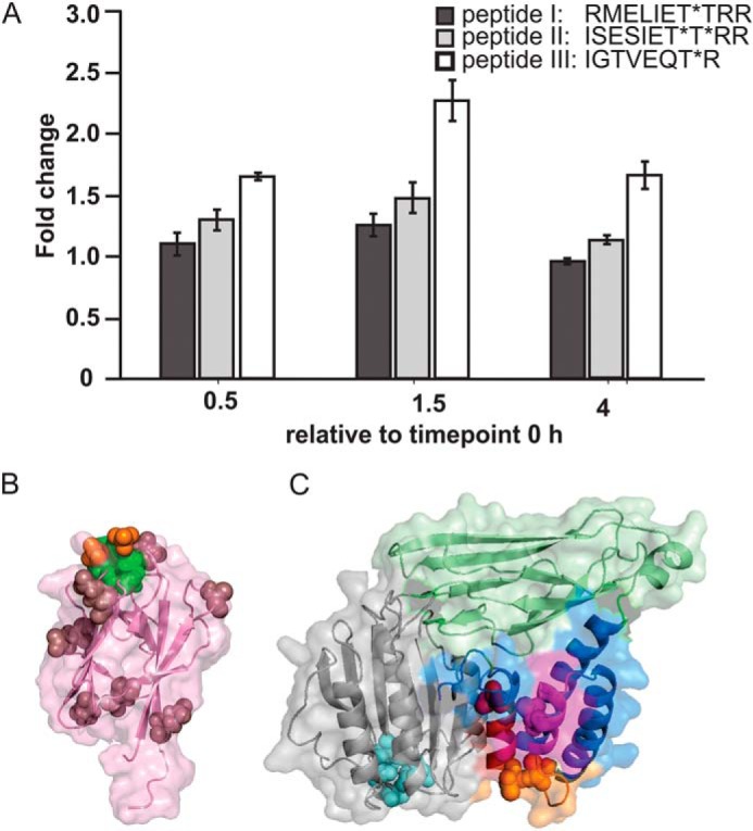
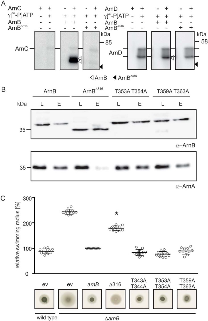
Phosphorylation-dependent interactions play crucial regulatory roles in all domains of life. Forkhead-associated (FHA) and von Willebrand type A (vWA) domains are involved in several phosphorylation-dependent processes of multiprotein complex assemblies. Although well-studied in eukaryotes and bacteria, the structural and functional contexts of these domains are not yet understood in Archaea. Here, we report the structural base for such an interacting pair of FHA and vWA domain-containing proteins, ArnA and ArnB, in the thermoacidophilic archaeon Sulfolobus acidocaldarius, where they act synergistically and negatively modulate motility. The structure of the FHA domain of ArnA at 1.75 Å resolution revealed that it belongs to the subclass of FHA domains, which recognizes double-pSer/pThr motifs. We also solved the 1.5 Å resolution crystal structure of the ArnB paralog vWA2, disclosing a complex topology comprising the vWA domain, a β-sandwich fold, and a C-terminal helix bundle. We further show that ArnA binds to the C terminus of ArnB, which harbors all the phosphorylation sites identified to date and is important for the function of ArnB in archaellum regulation. We also observed that expression levels of the archaellum components in response to changes in nutrient conditions are independent of changes in ArnA and ArnB levels and that a strong interaction between ArnA and ArnB observed during growth on rich medium sequentially diminishes after nutrient limitation. In summary, our findings unravel the structural features in ArnA and ArnB important for their interaction and functional archaellum expression and reveal how nutrient conditions affect this interaction.

Keywords: Archaea; cell motility; protein phosphorylation; signal transduction; transcription regulation.


Conflict of interest statement

The authors declare that they have no conflicts of interest with the contents of this article

Figures

Figure 1.
Figure 1.
Protein levels of ArnA, ArnB, and FlaB in S. acidocaldarius MW001 (WT) before and after nutrient depletion. A, cells were grown to an A600 of 0.4 (time point, 0 h) and then shifted to nutrient depleted medium (23, 24). Samples were taken after 0.5, 1.5, and 4 h of nutrient limitation (time points are depicted above the respective lanes), separated on an SDS-PAGE, and analyzed by Western blotting using ArnA (top row), ArnB (middle row), or FlaB (bottom row) specific primary antibodies. The analysis was repeated three times, and a representative Western blot is depicted. B, quantification of ArnA and ArnB levels. Levels of three independent experiments and two technical replicates were quantified relative to their intensities at time point 0 h.
Figure 2.
Figure 2.
Interaction of ArnA and ArnB in nutrient-rich and nutrient-depletion conditions. Pulldown experiments with MW390 (ΔarnB::arnB-Strep) were performed in exponentially grown cells (T = 0 h) and cells growing in nutrient depleted medium for 0.5, 1.5, and 4 h using Strep-Tactin–coated magnetic beads. A, ArnB and ArnA were detected in load (L) and elution (E) fractions using specific primary antibodies. Each pulldown and Western blotting was performed three times with independent replicates, and a representative blot is depicted. B, the ratio of ArnA/ArnB found in the elution fractions at the different time points was quantified and averaged for three independent experiments. The ArnA/ArnB ratio at the start of depletion was set to 1.
Figure 3.
Figure 3.
Crystal structure of the FHA domain from ArnA and cognate phospho-recognition module. A, overview of the FHA domain from ArnA (red) in cartoon and surface representation; the two molecules in spheres represent the sulfate ions in the structure. B, side view of the FHA domain. C, secondary structure topology of the FHA domain from ArnA; triangles indicate β-strands, and the directions of the tips refer to the orientation of the β-strand. D, comparison of the FHA domains from the archaeal ArnA (red) and the yeast's Rad53p–FHA1 from S. cerevisiae (cyan) (PDB code 1G6G); the structures superimpose with a r.m.s. deviation of 1.45 Å (61 Cα atoms). E, cognate phospho-recognition module in ArnA (panels 1–3). Primary and (panels 1, 4, and 5) putative secondary site for phospho-peptide binding (panels 1) are shown. Panel 2, the structure of ArnA (red) exhibits two sulfate ions at its surface; distances are displayed as dashed lines. Panel 3, structure of Rad53p–FHA1 from S. cerevisiae (cyan) (PDB code 1G6G) (9) in complex with a phosphothreonine peptide (blue). Panel 4, superimposition of ArnA and Rad53p–FHA1; at the position of the phosphate residue in the Rad53p–FHA1 structure, the ArnA structure exhibits the sulfate ion, suggesting a putative phospho-peptide–binding site at this position. The FHA domain of human PNK (pink) (PDB code 2W3O) binds a pThr-pSer peptide. pSer exhibits two rotamers in the structure. Panel 5, superimposition of ArnA and PNK-FHA.
Figure 4.
Figure 4.
Crystal structure of vWA2. A, overview of vWA2 in cartoon and surface representation. The vWA domain, the β-sandwich, and the helix bundle are displayed in gray, green, and blue, respectively. The N-terminal β-strands contributing to the β-sandwich are highlighted in light green. B, side view of vWA2. C, comparison between vWA2 (green) and the von Willebrand factor type A (cyan) from C. acidiphila (PDB code 4FX5) (r.m.s. deviation = 3.750 Å, 264 Cα atoms). D, secondary structure topology of vWA2. The coloration is based on A. Triangles indicate β-strands whose tips show the orientation of the β-strand, and circles represent α-helices. The β-sandwich exhibits a CnaA-like fold as the N2 domain of the adhesin Sgo0707 (48). E, MIDAS motif of vWA2 (top panel), the vWA-like gene product from C. acidiphila (middle panel), and from the α-subunit of the integrin CR3 (PDB code 1IDO) (bottom panel) (17).
Figure 5.
Figure 5.
The C terminus of ArnB is phosphorylated. A, changed abundances of phosphorylated peptides detected from ArnB during nutrient limitation. The data were compared with time = 0 h (before nutrient limitation). B, structure of ArnA highlighting the positions of surface-exposed glutamates (purple) and the site where a phosphothreonine peptide (red and green spheres) might bind. C, structural model of ArnB and localization of in vitro phosphorylated amino acids. The homology model was generated by SWISS-MODEL with vWA2 as template (42, 43) and shown in the same orientation as vWA2 in Fig. 4. The vWA domain is represented in gray, and the amino acids of its characteristic metal ion adhesion site (MIDAS) are in cyan. Phosphorylated residues identified by ITRAQ analysis and in a previous study (24) are depicted: red, pThr of peptide 1 (RMELIET*T*RR); magenta, pThr of peptide 2 (ISESIET*T*RR); and yellow, pThr of peptide 3 (IDT*VEQT*R).
Figure 6.
Figure 6.
The C terminus of ArnB is crucial for ArnA–ArnB interaction. A, in vitro phosphorylation of ArnB and ArnBΔ316 with the Ser/Thr kinase ArnC. ArnC autophosphorylates upon incubation with [γ-32P]ATP (left panels). Phosphotransfer from ArnC to WT ArnB (middle panels) but not ArnBΔ316 (right panels) was observed. The assays were repeated three times, and a representative phosphorimage is depicted. B, in vivo interaction of ArnA with Strep/His-ArnB, Strep/His-ArnBΔ316, and Strep-HisArnB phosphorylation ablative mutants. Western blotting analyses of the load (L) and elution (E) fractions obtained during pulldown analysis with Streptactin-coated magnetic beads were performed with ArnA and ArnB specific primary antibodies. C, swimming radius and motility assay of ΔarnB and ΔarnB complementation with the different ArnB mutants. Plasmid encoded arnBΔ316, arnB T343A/T344A, arnB T353A/T354A, and arnB T359A/T363A were used to complement the ΔarnB strain, and their swimming radii were subsequently compared with ΔarnB cells complemented with WT arnB. All strains were analyzed for their motility phenotype on semi-solid plates. After growth for 4 days at 75 °C, the swimming radii of the strains were calculated. Bars represent the mean relative swimming radius of three independent biological replicates with each six technical replicates normalized to the ΔarnB strain complemented with WT arnB in trans, which was set to 100%. Statistical significance was analyzed using a Student's t test (unequal variance, two-tailed) compared with the ΔarnB strain complemented with WT arnB. p values of <0.05 are indicated by asterisks.

Similar articles

Cited by

References

    1. Kennelly P. J. (2002) Protein kinases and protein phosphatases in prokaryotes: a genomic perspective. FEMS Microbiol. Lett. 206, 1–8 10.1111/j.1574-6968.2002.tb10978.x - DOI - PubMed
    1. Hofmann K., and Bucher P. (1995) The FHA domain: a putative nuclear signalling domain found in protein kinases and transcription factors. Trends Biochem. Sci. 20, 347–349 10.1016/S0968-0004(00)89072-6 - DOI - PubMed
    1. Mohammad D. H., and Yaffe M. B. (2009) 14–3-3 proteins, FHA domains and BRCT domains in the DNA damage response. DNA Repair (Amst.) 8, 1009–1017 10.1016/j.dnarep.2009.04.004 - DOI - PMC - PubMed
    1. Durocher D., and Jackson S. P. (2002) The FHA domain. FEBS Lett. 513, 58–66 10.1016/S0014-5793(01)03294-X - DOI - PubMed
    1. Niebisch A., Kabus A., Schultz C., Weil B., and Bott M. (2006) Corynebacterial protein kinase G controls 2-oxoglutarate dehydrogenase activity via the phosphorylation status of the OdhI protein. J. Biol. Chem. 281, 12300–12307 10.1074/jbc.M512515200 - DOI - PubMed

Publication types

MeSH terms