Case Reports
. 2019 Mar 21;12(3):e227128.
doi: 10.1136/bcr-2018-227128.

Image-guided volumetric modulated arc therapy (IG-VMAT) for unresectable ACC of the trachea: a feasible curative option

Affiliations
Case Reports

Image-guided volumetric modulated arc therapy (IG-VMAT) for unresectable ACC of the trachea: a feasible curative option

Rahul Lal Chowdhary et al. BMJ Case Rep. .

Abstract

A middle-aged man presented with progressively worsening breathlessness and non-productive cough for the last 3 months. On examination, his breathing was stridulous and air entry was decreased bilaterally. He underwent emergency fibre-optic bronchoscopy, which revealed a tracheal growth causing luminal narrowing, and after tumour debulking, he improved symptomatically. Histopathological evaluation of the specimen revealed an adenoid cystic carcinoma of the trachea, and systemic evaluation revealed metastatic dissemination. Systemic molecular-targeted therapy was initiated (gefitinib and later imatinib mesylate) and continued for 5 years, in view of stable disease on periodic follow-up. He subsequently presented with breathlessness again, which was managed with an emergency tracheostomy. In view of stable systemic disease and local progression only, he received definitive radiotherapy with image-guided volumetric modulated arc therapy, which resulted in a complete radiological response. The patient has been disease-free for the last 9 months.

Keywords: radiotherapy; respiratory cancer; tyrosine kinase inhibitor.


Conflict of interest statement

Competing interests: None declared.

Figures
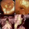
Figure 1
Figure 1
Bronchoscopy and 18FDG PET-CT images at the time of initial presentation. (A, B) Bronchoscopy images above and below the level of vocal cords, demonstrating an exophytic ulceroproliferative growth arising from the posterior wall of the trachea with consequent luminal narrowing. (C, D) PET-CT images in the axial and sagittal plane demonstrating the tracheal tumour (after debulking and delineated in red contour) abutting the anterior wall of the oesophagus and projecting into the tracheal lumen. 18FDG PET-CT, 18fluoro-deoxyglucose positron emission tomography.
Figure 2
Figure 2
Photomicrographs of biopsy specimen. (A,B) ACC with tubular and cribriform architecture lying beneath the mucosa of the trachea (A: H&E, ×100; B: H& E, ×200). (C,D) Section showing ACC abutting the tracheal cartilage (C: H&E, x100; D: H&E, x200). ACC,
Figure 3
Figure 3
Three-dimensional model view of the patient’s upper aerodigestive tract and tumour topography created with a combination of manual high-resolution delineation and semi-automatic Hounsfield Unit-based delineation. (A,B) Top down and lateral view, demonstrating the tumour (red) causing luminal narrowing of the trachea (orange) and abutting the oesophagus (light green). Also, note the invasion of the tumour beyond the confines of the trachea laterally. Laryngeal framework is shown in translucent blue and spinal column in light brown.
Figure 4
Figure 4
Diagnostic CT, bronchoscopy and treatment planning images. (A,B) Axial and sagittal images from the diagnostic CT done after emergency tracheostomy. The tumour is seen invading beyond the confines of the trachea with loss of fat planes with the oesophagus. (C,D) Bronchoscopy images (above and below the level of vocal cords) demonstrating an exophytic ulceroproliferative growth arising from the posterior wall of the trachea. (E,F) Dose colour wash in axial and sagittal planes (with lower limit set to 95% of 60 Gy) demonstrating conformal coverage of the PTV (red contour) and GTV (blue contour). GTV, gross tumour volume; PTV, planning target volume.
Figure 5
Figure 5
Post-RT CT images performed at 12 weeks. (A, B) Axial and sagittal images demonstrating complete response of the tracheal tumour with residual thickening at the posterior wall of the trachea. RT, radiotherapy.

Similar articles

References

    1. Honings J, van Dijck JA, Verhagen AF, et al. . Incidence and treatment of tracheal cancer: a nationwide study in the Netherlands. Ann Surg Oncol 2007;14:968–76. 10.1245/s10434-006-9229-z - DOI - PubMed
    1. Manninen MP, Antila PJ, Pukander JS, et al. . Occurrence of tracheal carcinoma in Finland. Acta Otolaryngol 1991;111:1162–9. 10.3109/00016489109100772 - DOI - PubMed
    1. Licht PB, Friis S, Pettersson G. Tracheal cancer in Denmark: a nationwide study. Eur J Cardiothorac Surg 2001;19:339–45. 10.1016/S1010-7940(01)00597-8 - DOI - PubMed
    1. Gelder CM, Hetzel MR. Primary tracheal tumours: a national survey. Thorax 1993;48:688–92. 10.1136/thx.48.7.688 - DOI - PMC - PubMed
    1. Bhattacharyya N. Contemporary staging and prognosis for primary tracheal malignancies: a population-based analysis. Otolaryngol Head Neck Surg 2004;131:639–42. 10.1016/j.otohns.2004.05.018 - DOI - PubMed

Publication types