. 2019 May 16;85(11):e02670-18.
doi: 10.1128/AEM.02670-18. Print 2019 Jun 1.

Biofilm-Constructing Variants of Paraburkholderia phytofirmans PsJN Outcompete the Wild-Type Form in Free-Living and Static Conditions but Not In Planta

Affiliations

Biofilm-Constructing Variants of Paraburkholderia phytofirmans PsJN Outcompete the Wild-Type Form in Free-Living and Static Conditions but Not In Planta

Marine Rondeau et al. Appl Environ Microbiol. .

Abstract

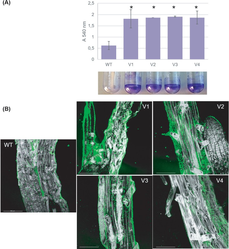
Members of the genus Burkholderia colonize diverse ecological niches. Among the plant-associated strains, Paraburkholderia phytofirmans PsJN is an endophyte with a broad host range. In a spatially structured environment (unshaken broth cultures), biofilm-constructing specialists of P. phytofirmans PsJN colonizing the air-liquid interface arose at high frequency. In addition to forming a robust biofilm in vitro and in planta on Arabidopsis roots, those mucoid phenotypic variants display a reduced swimming ability and modulate the expression of several microbe-associated molecular patterns (MAMPs), including exopolysaccharides (EPS), flagellin, and GroEL. Interestingly, the variants induce low PR1 and PDF1.2 expression compared to that of the parental strain, suggesting a possible evasion of plant host immunity. We further demonstrated that switching from the planktonic to the sessile form did not involve quorum-sensing genes but arose from spontaneous mutations in two genes belonging to an iron-sulfur cluster: hscA (encoding a cochaperone protein) and iscS (encoding a cysteine desulfurase). A mutational approach validated the implication of these two genes in the appearance of variants. We showed for the first time that in a heterogeneous environment, P. phytofirmans strain PsJN is able to rapidly diversify and coexpress a variant that outcompete the wild-type form in free-living and static conditions but not in plantaIMPORTANCEParaburkholderia phytofirmans strain PsJN is a well-studied plant-associated bacterium known to induce resistance against biotic and abiotic stresses. In this work, we described the spontaneous appearance of mucoid variants in PsJN from static cultures. We showed that the conversion from the wild-type (WT) form to variants (V) correlates with an overproduction of EPS, an enhanced ability to form biofilm in vitro and in planta, and a reduced swimming motility. Our results revealed also that these phenotypes are in part associated with spontaneous mutations in an iron-sulfur cluster. Overall, the data provided here allow a better understanding of the adaptive mechanisms likely developed by P. phytofirmans PsJN in a heterogeneous environment.

Keywords: Paraburkholderia phytofirmans; biofilm; competition; iron-sulfur cluster; plant defense; static cultures.


Figures

FIG 1
FIG 1
Emergence of P. phytofirmans PsJN variants from unshaken broth cultures. A single ancestral “WT” cell was propagated in 10 ml of King B medium at 28°C in a 50-ml microcosm without shaking for 8 days to produce a spatially heterogeneous environment. After 8 days, the culture was plated and showed substantial phenotypic diversity: a typical flat beige wild-type (WT) colony and an alternate phenotype, a mucoid white colony called variant (V).
FIG 2
FIG 2
Biofilm formation on an abiotic surface and in planta. (A) Biofilms were quantified by crystal violet staining after 24 h of static incubation at 28°C. A graphical representation of the optical density readings (OD540) derived from methanol elution of the crystal violet stain is shown. A partial F test was performed to determine if there were strain-specific differences in the level of biofilm formation (*, P < 0.05). (B) A. thaliana root attachment by P. phytofirmans PsJN::GFP 4 days after inoculation. Scanning confocal laser microscopy allowed visualization of green fluorescence emitted by the bacteria. Bars, = 100 μm.
FIG 3
FIG 3
EPS production and motility in the wild type and variants. (A) EPS production by wild-type P. phytofirmans (WT) and variants (V1 to V4). EPS production was tested on mannitol medium containing 0.02% yeast extract. (B) Swimming motility (on LB plates with 0.2% agar without salt and supplemented with 0.1% Casamino Acids) was measured after 30 h of incubation. Error bars indicate standard errors (SE). Asterisks indicate a significant difference between the WT and others strains (****, P < 0.0001). (C) Negative-stain transmission electron micrographs (TEM) of WT P. phytofirmans PsJN and variants (V1 to V4). The strains were grown to mid-log phase in KB medium, transferred onto Formvar-coated nickel grids, and stained with phosphotungstic acid. Bars, = 0.5 μm.
FIG 4
FIG 4
Defense gene expression in A. thaliana inoculated or not with P. phytofirmans PsJN (WT or V) or E. coli. Transcript accumulation of the PR1 or PDF1.2 gene was determined by quantitative real-time PCR at 24 h postinoculation with bacteria. Gene transcript levels were normalized using two reference genes (UBQ5 and EF1a) as internal controls. Results are expressed as the fold increase in transcript level compared to that in control plants. Values shown are means ± standard deviations (SD) from two independent repetitions (each repetition was done in duplicates).
FIG 5
FIG 5
Genetic organization of the P. phytofirmans PsJN Iron Sulfur Cluster (ISC) system. iscR transcriptional regulator, Rrf2 family; iron-sulfur cluster assembly transcription factor IscR (WP_012433568.1); iscS cysteine desulfurase (WP_012433567.1); iscU FeS cluster assembly scaffold IscU (WP_012433566.1); iscA iron-sulfur cluster assembly protein IscA (WP_012433565.1); hscB chaperone protein HscB (WP_012433564.1); hscA cochaperone protein HscA (WP_012433563.1); fdx ferredoxin 2Fe-2S type (WP_012433562.1); iscX FeS assembly protein IscX (WP_007181290.1).
FIG 6
FIG 6
Characterization of ΔhscA and ΔiscS mutants. (A to C) Motility (A), EPS production (B), and biofilm (C) of ΔhscA and ΔiscS mutants compared to WT cells. (D) Defense gene expression in A. thaliana inoculated or not with P. phytofirmans PsJN (WT or mutants) or E. coli.
FIG 7
FIG 7
In vitro bacterial competition. A mixture of dsRed-tagged WT P. phytofirmans and GFP-tagged P. phytofirmans V (A) or of GFP-tagged WT P. phytofirmans and dsRed-tagged P. phytofirmans mutant (B) was tested for competition after 72 h on plates and 24 h in shaking and static cultures. The percentage of CFU of the two competing strains in the inoculum and recovered after 72 h (on plate) or 24 h (shaking and static) of contact is shown. Error bars indicate standard deviations (SD). At least 3 independent replicates were analyzed.
FIG 8
FIG 8
In planta bacterial competition. A mixture of dsRed-tagged WT P. phytofirmans and GFP-tagged P. phytofirmans V (A) or of GFP-tagged WT P. phytofirmans and P. phytofirmans mutant dsRed-tagged (B) was tested for in planta competition at 2, 6, and 12 days after inoculation (dai). The percentage of CFU of the two competing strains in the inoculum and recovered at 2, 6, and 12 dai (in planta) is shown. Error bars indicate standard deviations (SD). At least 2 independent repetitions (each in duplicates) were analyzed.

References

    1. Coenye T, Vandamme P. 2003. Diversity and significance of Burkholderia species occupying diverse ecological niches. Environ Microbiol 5:719–729. doi:10.1046/j.1462-2920.2003.00471.x. - DOI - PubMed
    1. Compant S, Nowak J, Coenye T, Clément C, Ait Barka E. 2008. Diversity and occurrence of Burkholderia spp. in the natural environment. FEMS Microbiol Rev 32:607–626. doi:10.1111/j.1574-6976.2008.00113.x. - DOI - PubMed
    1. Eberl L, Vandamme P. 2016. Members of the genus Burkholderia: good and bad guys. F1000Res 5:F1000 Faculty Rev-1007. doi:10.12688/f1000research.8221.1. - DOI - PMC - PubMed
    1. Chain PS, Denef VJ, Konstantinidis KT, Vergez LM, Agulló L, Reyes VL, Hauser L, Córdova M, Gómez L, González M, Land M, Lao V, Larimer F, LiPuma JJ, Mahenthiralingam E, Malfatti SA, Marx CJ, Parnell JJ, Ramette A, Richardson P, Seeger M, Smith D, Spilker T, Sul WJ, Tsoi TV, Ulrich LE, Zhulin IB, Tiedje JM. 2006. Burkholderia xenovorans LB400 harbors a multi-replicon, 9.73-Mbp genome shaped for versatility. Proc Natl Acad Sci U S A 103:15280–15287. doi:10.1073/pnas.0606924103. - DOI - PMC - PubMed
    1. Sawana A, Adeolu M, Gupta RS. 2014. Molecular signatures and phylogenomic analysis of the genus Burkholderia: proposal for division of this genus into the emended genus Burkholderia containing pathogenic organisms and a new genus Paraburkholderia gen. nov. harboring environmental species. Front Genet 5:429. doi:10.3389/fgene.2014.00429. - DOI - PMC - PubMed

Publication types

Supplementary concepts