. 2019 May 29;39(22):4375-4386.
doi: 10.1523/JNEUROSCI.2497-18.2019. Epub 2019 Mar 22.

The Neural Dynamics of Novel Scene Imagery

Affiliations

The Neural Dynamics of Novel Scene Imagery

Daniel N Barry et al. J Neurosci. .

Abstract

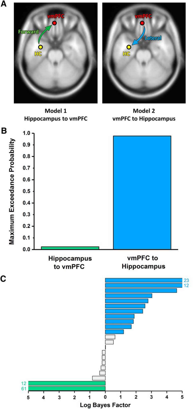
Retrieval of long-term episodic memories is characterized by synchronized neural activity between hippocampus and ventromedial prefrontal cortex (vmPFC), with additional evidence that vmPFC activity leads that of the hippocampus. It has been proposed that the mental generation of scene imagery is a crucial component of episodic memory processing. If this is the case, then a comparable interaction between the two brain regions should exist during the construction of novel scene imagery. To address this question, we leveraged the high temporal resolution of MEG to investigate the construction of novel mental imagery. We tasked male and female humans with imagining scenes and single isolated objects in response to one-word cues. We performed source-level power, coherence, and causality analyses to characterize the underlying interregional interactions. Both scene and object imagination resulted in theta power changes in the anterior hippocampus. However, higher theta coherence was observed between the hippocampus and vmPFC in the scene compared with the object condition. This interregional theta coherence also predicted whether imagined scenes were subsequently remembered. Dynamic causal modeling of this interaction revealed that vmPFC drove activity in hippocampus during novel scene construction. Additionally, theta power changes in the vmPFC preceded those observed in the hippocampus. These results constitute the first evidence in humans that episodic memory retrieval and scene imagination rely on similar vmPFC-hippocampus neural dynamics. Furthermore, they provide support for theories emphasizing similarities between both cognitive processes and perspectives that propose the vmPFC guides the construction of context-relevant representations in the hippocampus.SIGNIFICANCE STATEMENT Episodic memory retrieval is characterized by a dialog between hippocampus and ventromedial prefrontal cortex (vmPFC). It has been proposed that the mental generation of scene imagery is a crucial component of episodic memory processing. An ensuing prediction would be of a comparable interaction between the two brain regions during the construction of novel scene imagery. Here, we leveraged the high temporal resolution of MEG and combined it with a scene imagination task. We found that a hippocampal-vmPFC dialog existed and that it took the form of vmPFC driving the hippocampus. We conclude that episodic memory and scene imagination share fundamental neural dynamics and the process of constructing vivid, spatially coherent, contextually appropriate scene imagery is strongly modulated by vmPFC.

Keywords: MEG; connectivity; hippocampus; scene construction; temporal dynamics; vmPFC.


Figures

Figure 1.
Figure 1.
Trial structure. The task period selected for analysis is highlighted.
Figure 2.
Figure 2.
MEG source reconstruction of theta (4–8 Hz) and alpha (9–12 Hz) power changes during mental imagery (scenes and objects) compared with the baseline condition. The black circles represent the peak location of theta power (A) and alpha power (B) changes in anterior hippocampus used for subsequent connectivity analyses. Images are FWE thresholded at p < 0.001 and superimposed on the Montreal Neurological Institute 152 T1 image.
Figure 3.
Figure 3.
Gradient of theta power change along the hippocampal axis during mental imagery. The magnitude of theta power change increased significantly from the posterior to anterior segments. This linear increase along the hippocampal axis was present in 15 of the 22 participants.
Figure 4.
Figure 4.
Brain areas displaying higher theta coherence with the left anterior hippocampus during scene imagination compared with object imagination. A, Fusiform and parahippocampal cortices showed higher coherence with the hippocampal source (black circle), display thresholded at p < 0.001 uncorrected, superimposed on the Montreal Neurological Institute 152 T1 image. B, The vmPFC, display thresholded at p < 0.005 uncorrected, also showed higher coherence with the hippocampal source (black circle). No areas showed higher theta coherence with the hippocampus for object over scene imagery. C, Coherence between the vmPFC and hippocampus was significantly higher for subsequently remembered than forgotten scenes. *p = 0.018. D, A similar trend was observed for scenes imagined in high versus low detail. †p = 0.058. Error bars indicate ± 1 SEM.
Figure 5.
Figure 5.
DCM of the interaction between the hippocampus and vmPFC. A, Two proposed models of effective connectivity between the coherent peaks in hippocampus and vmPFC. B, Results of Bayesian model comparison indicated a stronger influence of the vmPFC on hippocampal activity during scene imagination. C, Log Bayes factors for each participant. Blue bars indicate positive to strong evidence for vmPFC driving hippocampus, the model that most consistently fit across participants. Green bars represent the only two cases where evidence of the hippocampus driving vmPFC was observed. Gray bars represent the remaining participants where there was no conclusive evidence for either model. Where log Bayes factors exceeded five, bars are truncated and the exact values are displayed adjacently.
Figure 6.
Figure 6.
Temporal profile of theta power changes in the vmPFC and hippocampus during scene construction. Line segments in bold indicate periods of significant (p < 0.05 FWE corrected) changes in power relative to the pre-imagination period for both the vmPFC (red) and hippocampus (yellow). An initial peak in vmPFC activity was observed 310 ms following cue offset, consistent with a role for the vmPFC in driving hippocampal activity. Maximal activation of both regions occurred within a narrow temporal window (hippocampus: 806 ms, vmPFC: 899 ms). A peak of similar magnitude was evident in the vmPFC at a later stage in the imagination period (1752 ms).

References

    1. Aggleton JP, Wright NF, Rosene DL, Saunders RC (2015) Complementary patterns of direct amygdala and hippocampal projections to the macaque prefrontal cortex. Cereb Cortex 25:4351–4373. 10.1093/cercor/bhv019 - DOI - PMC - PubMed
    1. Backus AR, Schoffelen JM, Szebényi S, Hanslmayr S, Doeller CF (2016) Hippocampal-prefrontal theta oscillations support memory integration. Curr Biol 26:450–457. 10.1016/j.cub.2015.12.048 - DOI - PubMed
    1. Balota DA, Yap MJ, Cortese MJ, Hutchison KA, Kessler B, Loftis B, Neely JH, Nelson DL, Simpson GB, Treiman R (2007) The English lexicon project. Behav Res Methods 39:445–459. 10.3758/BF03193014 - DOI - PubMed
    1. Barnes GR, Hillebrand A, Fawcett IP, Singh KD (2004) Realistic spatial sampling for MEG beamformer images. Hum Brain Mapp 23:120–127. 10.1002/hbm.20047 - DOI - PMC - PubMed
    1. Barry DN, Chadwick MJ, Maguire EA (2018) Nonmonotonic recruitment of ventromedial prefrontal cortex during remote memory recall. PLoS Biol 16:e2005479. 10.1371/journal.pbio.2005479 - DOI - PMC - PubMed

Publication types

LinkOut - more resources