. 2019 Jun 5;39(23):4606-4623.
doi: 10.1523/JNEUROSCI.3069-18.2019. Epub 2019 Mar 22.

Astrocytic Epoxyeicosatrienoic Acid Signaling in the Medial Prefrontal Cortex Modulates Depressive-like Behaviors

Affiliations

Astrocytic Epoxyeicosatrienoic Acid Signaling in the Medial Prefrontal Cortex Modulates Depressive-like Behaviors

Wenchao Xiong et al. J Neurosci. .

Abstract

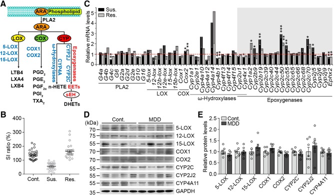
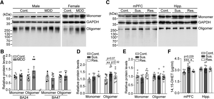
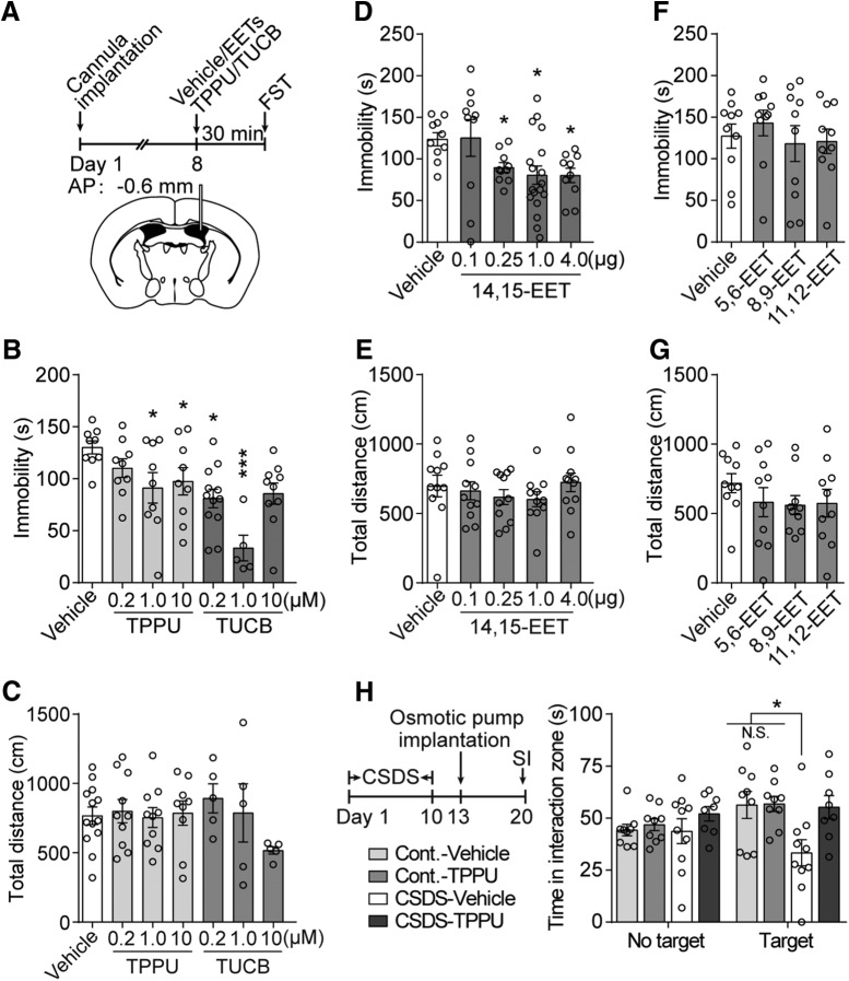
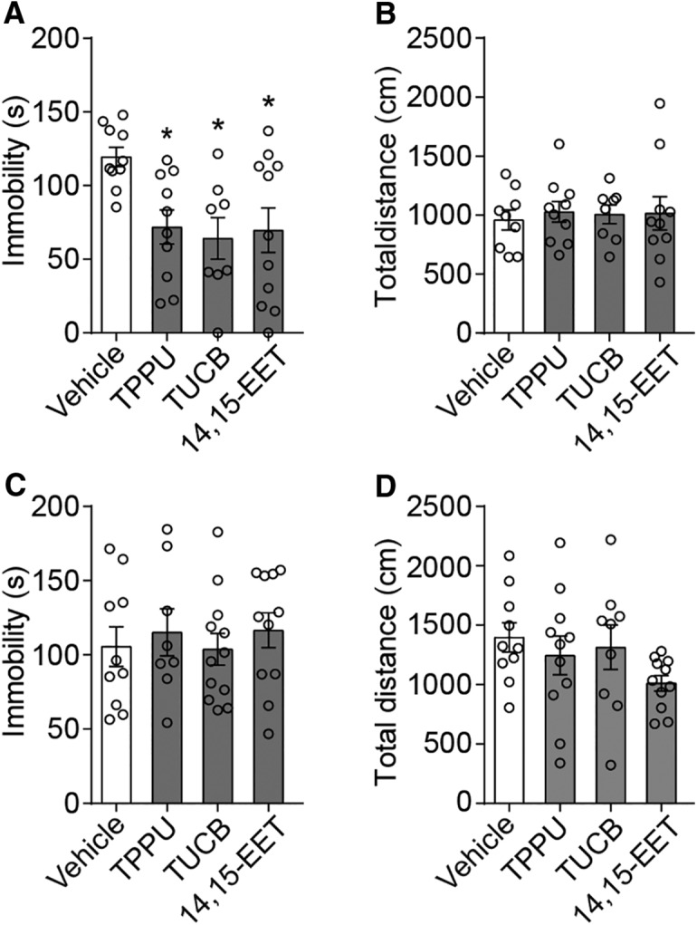
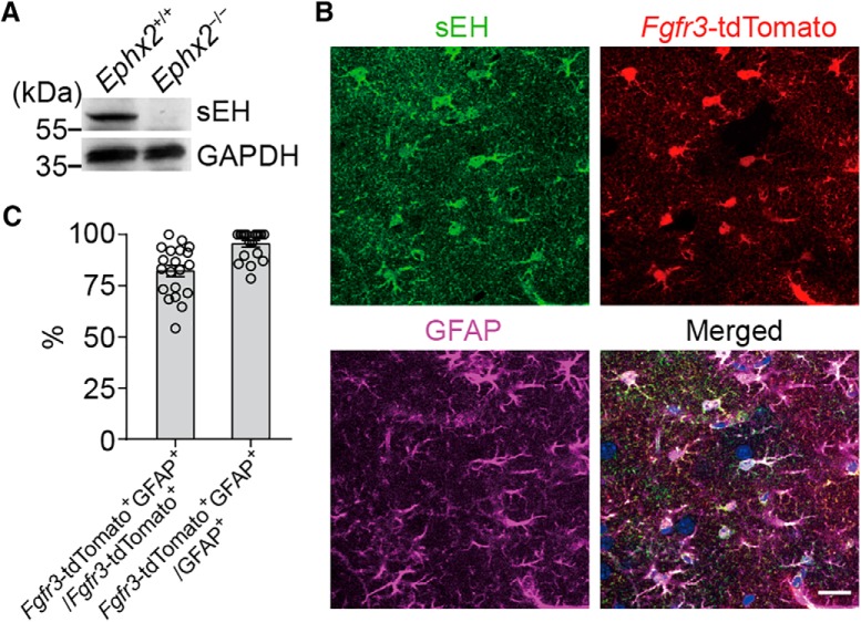
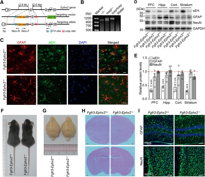
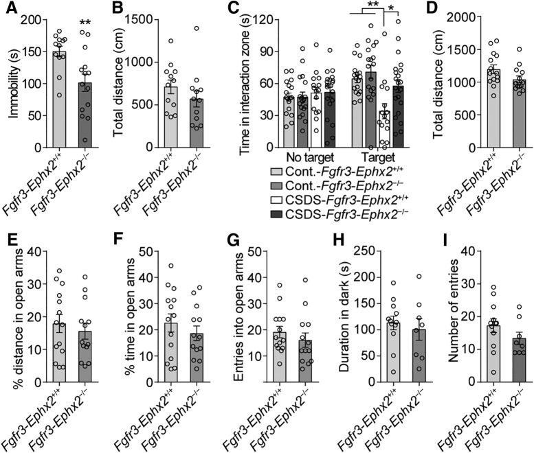
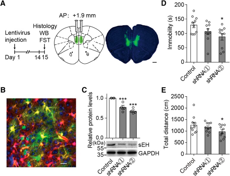
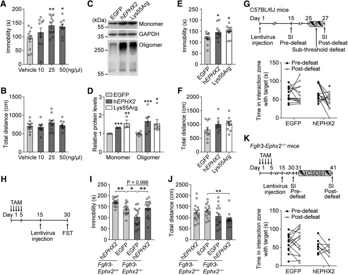
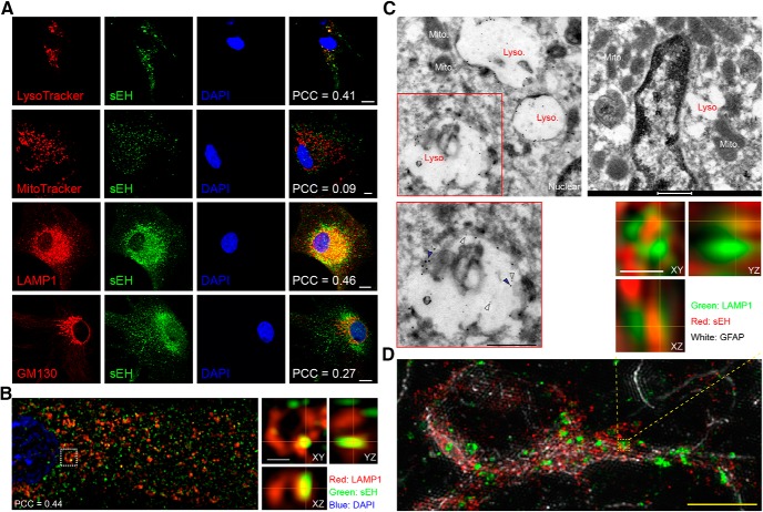
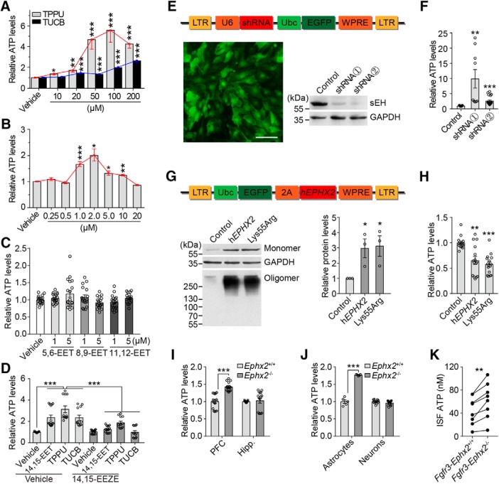
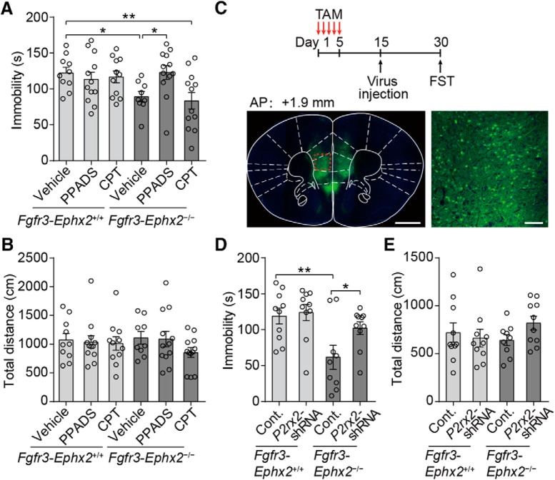
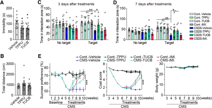
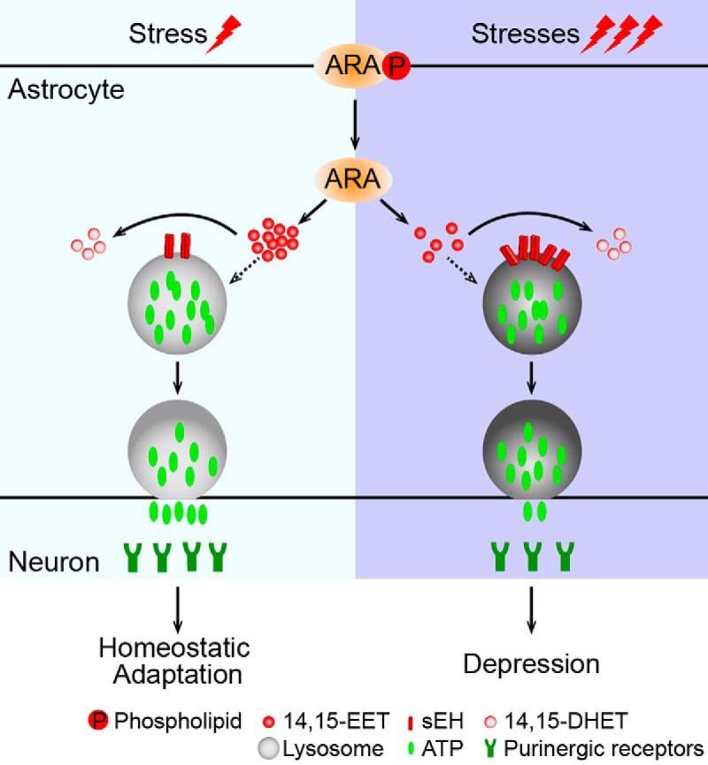
Major depressive disorder is the most common mental illness. Mounting evidence indicates that astrocytes play a crucial role in the pathophysiology of depression; however, the underlying molecular mechanisms remain elusive. Compared with other neuronal cell types, astrocytes are enriched for arachidonic acid metabolism. Herein, we observed brain-region-specific alterations of epoxyeicosatrienoic acid (EET) signaling, which is an arachidonic acid metabolic pathway, in both a mouse model of depression and postmortem samples from patients with depression. The enzymatic activity of soluble epoxide hydrolase (sEH), the key enzyme in EET signaling, was selectively increased in the mPFC of susceptible mice after chronic social defeated stress and was negatively correlated with the social interaction ratio, which is an indicator of depressive-like behavior. The specific deletion of Ephx2 (encode sEH) in adult astrocytes induced resilience to stress, whereas the impaired EET signaling in the mPFC evoked depressive-like behaviors in response to stress. sEH was mainly expressed on lysosomes of astrocytes. Using pharmacological and genetic approaches performed on C57BL/6J background adult male mice, we found that EET signaling modulated astrocytic ATP release in vitro and in vivo Moreover, astrocytic ATP release was required for the antidepressant-like effect of Ephx2 deletion in adult astrocytes. In addition, sEH inhibitors produced rapid antidepressant-like effects in multiple animal models of depression, including chronic social defeated stress and chronic mild stress. Together, our results highlight that EET signaling in astrocytes in the mPFC is essential for behavioral adaptation in response to psychiatric stress.SIGNIFICANCE STATEMENT Astrocytes, the most abundant glial cells of the brain, play a vital role in the pathophysiology of depression. Astrocytes secrete adenosine ATP, which modulates depressive-like behaviors. Notably, astrocytes are enriched for arachidonic acid metabolism. In the present study, we explored the hypothesis that epoxyeicosatrienoic acid signaling, an arachidonic acid metabolic pathway, modulates astrocytic ATP release and the expression of depressive-like behaviors. Our work demonstrated that epoxyeicosatrienoic acid signaling in astrocytes in the mPFC is essential for behavioral homeostatic adaptation in response to stress, and the extent of astrocyte functioning is greater than expected based on earlier reports.

Keywords: ATP; astrocytes; depression; epoxyeicosatrienoic acid signaling; medial prefrontal cortex; soluble epoxide hydrolase.


Figures

Figure 1.
Figure 1.
ARA metabolic pathways in the mouse model and patients with MDD. A, Schematic representation of the ARA metabolic pathway. PLA2 enzymes are crucial for transferring esterified ARA to free ARA for metabolism. Three members of the PLA2 superfamily have been implicated most strongly in eicosanoid production, including cytosolic calcium-dependent PLAs [group (G) 4a–c], cytosolic calcium-independent PLA2 (G6), and secreted PLA2 (G2a, b, e and G10). Adipose-specific PLA2 is G16. The free ARA can be converted to eicosanoids via three pathways: the LOX, COX, and CYP. B, Identification of susceptible and resilient mice following the CSDS paradigm. Mice were divided into different groups by the SI ratio. Cont., Control; Sus., susceptible; Res., resilient. C, mRNA levels (relative to the control, red dashed line) of gene profiles related to the ARA KEGG pathway in the mPFC of adult C57BL/6J mice after CSDS. For qPCR primers, see Figure 1-1. D, E, Western blots and quantification of LOXs, COXs, and CYPs in the BA24 of subjects with MDD and matched controls. Data are mean ± SEM. *p < 0.05, **p < 0.01, ***p < 0.001.
Figure 2.
Figure 2.
Dysfunction of EET signaling in human depression and mouse CSDS. A, Western blots showing the expression of sEH monomer and oligomer in the BA24 of patients with MDD and matched controls. B, Quantification of the Western blot results in BA24 and BA47. Hollow circle represents male. Solid circle represents female. C, Western blot analyses of sEH in the mPFC and hippocampus (Hipp.) after CSDS. D, E, Quantifications of sEH monomer and oligomer in the mPFC and hippocampus after CSDS. F, Measurement of the levels of 14,15-DHET hydrolyzed by sEH in the mPFC and hippocampus after CSDS. Cont., Control; Sus., susceptible; Res., resilient. Data are mean ± SEM. *p < 0.05, **p < 0.01, ***p < 0.001.
Figure 3.
Figure 3.
Antidepressant-like effects are induced by enhancement of EET signaling. A, Schematic representation of the experimental design for intracerebroventricular injections. B, Immobility of adult C57BL/6J mice injected with different dosages of TPPU, TUCB, or vehicle in the FST. C, No effect on spontaneous locomotor activity was observed following an intracerebroventricular infusion of different dosages of TPPU, TUCB, or vehicle. D, Immobility of adult C57BL/6J mice injected with different dosages of 14,15-EET in the FST. E, Infusions of 14,15-EET had no effect on locomotion. F, G, The intracerebroventricular infusions of 5,6-, 8,9-, or 11,12-EET did not produce antidepressant-like effects in the FST. H, Schematic paradigm and the antidepressant-like effect of TPPU (intracerebroventricular) in the CSDS model. Data are mean ± SEM. *p < 0.05, ***p < 0.001. N.S., not significant.
Figure 4.
Figure 4.
Enhancement of EET signaling in the mPFC produces antidepressant-like effects. A, Effect of mPFC infusion of TPPU (1 μm), TUCB (0.1 μm), 14,15-EET (1 μg), or vehicle on immobility in the FST. B, mPFC infusions of TPPU (1 μm), TUCB (0.1 μm), or 14,15-EET (1 μg) did not affect locomotor activity compared with the controls. C, D, Hippocampal infusions of TPPU (1 μm), TUCB (0.1 μm), or 14,15-EET (1 μg) could not induce antidepressant-like effects in the FST. Data are mean ± SEM. *p < 0.05.
Figure 5.
Figure 5.
sEH is mainly expressed in astrocytes in the mPFC. A, Western blot analysis of sEH in the brain of Ephx2−/− and control littermates. B, Brain slices (40 μm) were prepared from adult Fgfr3-tdTomato mice and stained with anti-sEH (green) and anti-GFAP (magenta). Scale bar, 20 μm. C, The statistical analysis indicates that >80% of tdTomato-positive cells in the mPFC colocalized with GFAP and most GFAP-positive cells were tdTomato-positive cells in the mPFC.
Figure 6.
Figure 6.
Generation of Fgfr3-Ephx2−/− mice. A, Schematic diagram of Ephx2loxp/loxp allele generation: the conditional Ephx2 allele has loxp sites flanking exon 2 and generates an out-of-frame mutation after Cre-mediated recombination. B, PCR genotyping of Ephx2loxp/loxp mice. C, Representative confocal images showing coexpression of sEH (green) in GFAP-positive astrocytes (red) in the mPFC of adult Fgfr3-Ephx2−/− mice and littermate controls. Scale bar, 20 μm. D, E, Western blot analysis of the levels of sEH, GFAP, and NeuN in the PFC, hippocampus (Hipp.), cerebral cortex (Cort.), and striatum of adult Fgfr3-Ephx2−/− mice and littermate controls. The levels of sEH were decreased in the PFC, hippocampus, cerebral cortex, and striatum by 66%, 58%, 57%, and 72%, respectively. F, Representative images of adult Fgfr3-Ephx2−/− and littermate control animals. G, Gross appearance of the brain of Fgfr3-Ephx2−/− and littermate control mice. H, H&E-stained coronal sections around the PFC (top) and hippocampus (bottom) of the brains of Fgfr3-Ephx2−/− and littermate control mice. Scale bar, 500 μm. I, Immunofluorescence for GFAP (green) in the hippocampus and NeuN (green) in the mPFC of Fgfr3-Ephx2−/− and littermate control mice. Scale bar, 20 μm. **p < 0.01, ***p < 0.001.
Figure 7.
Figure 7.
Deletion of Ephx2 in adult astrocytes induces resilience to stress. A, Antidepressant-like effect of Ephx2 deletion in adult astrocytes in the FST. B, Fgfr3-Ephx2−/− and littermate control mice were detected in the open field test. C, Fgfr3-Ephx2−/− mice were less susceptible to social defeat stress. D–G, Behavioral responses of Fgfr3-Ephx2−/− mice and littermate controls in the elevated plus maze test. H, I, Animals were examined in the light-dark box test. Data are mean ± SEM. *p < 0.05, **p < 0.01.
Figure 8.
Figure 8.
Enhancement of EET signaling in adult astrocytes in the mPFC produces antidepressant-like effects. A, Experimental timeline (left) and bilateral injection sites in the mPFC (middle) with an image of EGFP expression 2 weeks after injection (right). Scale bar, 0.5 mm. B, Confocal images of the mPFC of adult Fgfr3-tdTomato mice 14 d after viral injection. Green represents pLenti-EGFP. Red represents tdTomato. Blue represents DAPI. Scale bar, 20 μm. C, Levels of sEH in the mPFC of adult C57BL/6J mice injected with pLenti-shRNAs. D, Immobility of adult C57BL/6J mice injected with pLenti-Ephx2-shRNAs or control shRNA. E, Spontaneous locomotor activity of mice with bilateral mPFC infusions of pLenti-shRNAs. Data are mean ± SEM. *p < 0.05, ***p < 0.001.
Figure 9.
Figure 9.
Impaired EET signaling in the mPFC induces a depressive-like phenotype. A, mPFC infusion of 14,15-EEZE increased the duration of immobility in the FST at the optimal concentrations. B, mPFC infusions of 14,15-EEZE had no effect on locomotion. C, D, Western blots and quantification of sEH monomer and oligomer in the mPFC of mice injected with pLenti-EGFP (control), pLenti-hEPHX2 (hEPHX2), or pLenti-Lys55Arg (Lys55Arg). E, F, Immobility of adult C57BL/6J mice injected with pLenti-hEPHX2, pLenti-Lys55Arg, or control virus in the FST. G, Schematic of the overexpression of hEPHX2 or control EGFP followed by subthreshold defeat (top), and behavioral analyses were conducted before (Pre-defeat) and after (Post-defeat) subthreshold defeat (bottom). H, Schematic of TAM-induced Ephx2 deletion in astrocytes, followed by overexpression of hEPHX2 or control EGFP in the mPFC in the FST. I, J, Immobility time (I) and total distance (J) of Fgfr3-Ephx2−/− mice and littermate controls injected with pLenti-hEPHX2 or control virus. K, Schematic of Ephx2 deletion followed by hEPHX2 overexpression in the mPFC in the CSDS paradigm (top), and social interaction measured before and after CSDS (bottom). Data are mean ± SEM. *p < 0.05, **p < 0.01, ***p < 0.001.
Figure 10.
Figure 10.
sEH is mainly located on lysosomes in astrocytes. A, Confocal images of astrocytes showing sEH (green) colabeling with different organelle markers (red). Scale bar, 10 μm. PCC, Pearson's correlation coefficient. B, SIM images showing coexpression of sEH (green) and LAMP1 (red) in cultured astrocytes. Scale bar, 0.5 μm. C, Immuno-TEM image showing coexpression of sEH (5 nm gold, feint arrows) and LAMP1 (10 nm gold, solid arrows) in cultured astrocytes. Scale bar, 0.5 μm. Lyso., Lysosome; Mito., mitochondria. D, Triple-IF staining of brain slices of adult C57BL/6J mice. SIM images showing coexpression of sEH (red) and LAMP1 (green) in astrocytes (white) in the mPFC. Scale bars: Top right, 0.5 μm; Bottom, 5 μm.
Figure 11.
Figure 11.
EET signaling modulates astrocytic ATP release. A, ATP measurements showing ATP levels in the medium of cultured astrocytes 5 min after treatment with different dosages of TPPU and TUCB. B, ATP levels in the medium of cultured astrocytes after 14,15-EET treatments. C, Five-minute treatments with 5,6-, 8,9-, and 11,12-EET (1 μm) had no effect on ATP release from cultured astrocytes. D, Effects of 14,15-EET (1 μm), TPPU (100 μm), and TUCB (50 μm) on astrocytic ATP release were attenuated by pretreatment with 14,15-EEZE. E, Schematic of the lentiviral vector encoding shRNA (top) and an image of EGFP expression (bottom left; scale bar, 10 μm) with Western blot analysis of sEH expression in cultured astrocytes (bottom right). LTR, Lentiviral long terminal repeat; U6, U6 promoter; UbC, ubiquitin C promoter; WPRE, woodchuck hepatitis post-transcriptional regulatory element. F, ATP levels in the medium of cultured astrocytes transfected with pLenti-shRNAs. G, Schematic of the lentiviral vector encoding hEPHX2 (top). Western blot and quantification showing levels of sEH oligomer in cultured astrocytes after overexpression of hEPHX2 and Lys55Arg. H, Amount of ATP in the medium of cultured astrocytes overexpressing pLenti-EGFP, hEPHX2, or Lys55Arg 72 h after transfection. I, ATP levels in the ACSF medium incubated with slices of the PFC or hippocampus (Hipp.) isolated from adult Ephx2−/− mice and littermate controls. J, ATP levels in the medium of cultured astrocytes or neurons isolated from neonatal Ephx2−/− mice and control animals. K, In vivo microdialysis assay showing ATP levels in the interstitial fluid (ISF) in the mPFC of adult Fgfr3-Ephx2−/− and control animals. All in vitro studies were repeated two or three times. Data are mean ± SEM. *p < 0.05, **p < 0.01, ***p < 0.001.
Figure 12.
Figure 12.
P2X2 receptors in the mPFC mediate the antidepressant-like effect induced by Ephx2 deletion in adult astrocytes. A, Effects of mPFC infusions of PPADS or CPT on the antidepressant phenotype observed in Fgfr3-Ephx2−/− mice. B, mPFC infusions of PPADS or CPT had no effect on locomotion. C, Top, Experimental time schedule. Bottom, Bilateral injection sites in the mPFC with an image of EGFP expression. Scale bars: Left, 1 mm; Right, 100 μm. D, Effects of mPFC microinjection with AAV-P2rx2-shRNA on the antidepressant phenotype observed in Fgfr3-Ephx2−/− mice. E, mPFC microinjection with AAV-P2rx2-shRNA had no effect on locomotion. Data are mean ± SEM. *p < 0.05, **p < 0.01.
Figure 13.
Figure 13.
sEHIs produce rapid antidepressant-like effects. A, The effects of TPPU, TUCB, and vehicle in the FST when mice were examined 1 h after the injection (i.p.). B, Treatments with sEHIs had no effect on locomotion. C, D, CSDS paradigm. Avoidance behaviors of the mice after 3 or 7 d treatment with vehicle, IMI, TPPU, or TUCB (i.p.). E, CMS paradigm. Measurements of sucrose preference (left), the physical state of the coat (middle) and body weight (right), showing baseline followed by poststress measurements conducted each week for the mice treated with vehicle, TPPU, TUCB, or IMI (i.p.). Data are mean ± SEM. *p < 0.05, **p < 0.01, ***p < 0.001. N.S., not significant.
Figure 14.
Figure 14.
EET signaling in astrocytes in the mPFC is essential for behavioral adaptation. Left, Behavioral adaptation (for details, see Discussion). Right, Chronic stress increased the activity of sEH and, thus, disrupted EET signaling in astrocytes, which induced a deficiency of astrocytic ATP release and induced depressive-like behaviors.

Similar articles

Cited by

References

    1. Allen NJ, Eroglu C (2017) Cell biology of astrocyte-synapse interactions. Neuron 96:697–708. 10.1016/j.neuron.2017.09.056 - DOI - PMC - PubMed
    1. An H, Cho DW, Lee SE, Yang YS, Han SC, Lee CJ (2016) Differential cellular tropism of lentivirus and adeno-associated virus in the brain of cynomolgus monkey. Exp Neurobiol 25:48–54. 10.5607/en.2016.25.1.48 - DOI - PMC - PubMed
    1. Banasr M, Duman RS (2008) Glial loss in the prefrontal cortex is sufficient to induce depressive-like behaviors. Biol Psychiatry 64:863–870. 10.1016/j.biopsych.2008.06.008 - DOI - PMC - PubMed
    1. Banasr M, Chowdhury GM, Terwilliger R, Newton SS, Duman RS, Behar KL, Sanacora G (2010) Glial pathology in an animal model of depression: reversal of stress-induced cellular, metabolic and behavioral deficits by the glutamate-modulating drug riluzole. Mol Psychiatry 15:501–511. 10.1038/mp.2008.106 - DOI - PMC - PubMed
    1. Banasr M, Dwyer JM, Duman RS (2011) Cell atrophy and loss in depression: reversal by antidepressant treatment. Curr Opin Cell Biol 23:730–737. 10.1016/j.ceb.2011.09.002 - DOI - PMC - PubMed

Publication types

MeSH terms

LinkOut - more resources