. 2019 Mar 22;10(1):1355.
doi: 10.1038/s41467-019-09377-6.

Phototrophic extracellular electron uptake is linked to carbon dioxide fixation in the bacterium Rhodopseudomonas palustris

Affiliations

Phototrophic extracellular electron uptake is linked to carbon dioxide fixation in the bacterium Rhodopseudomonas palustris

Michael S Guzman et al. Nat Commun. .

Abstract

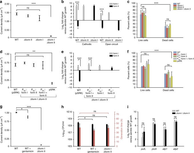
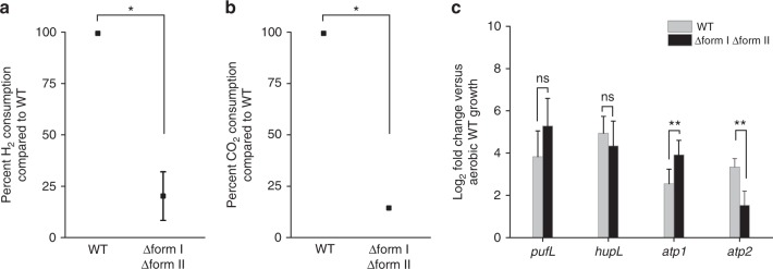
Extracellular electron uptake (EEU) is the ability of microbes to take up electrons from solid-phase conductive substances such as metal oxides. EEU is performed by prevalent phototrophic bacterial genera, but the electron transfer pathways and the physiological electron sinks are poorly understood. Here we show that electrons enter the photosynthetic electron transport chain during EEU in the phototrophic bacterium Rhodopseudomonas palustris TIE-1. Cathodic electron flow is also correlated with a highly reducing intracellular redox environment. We show that reducing equivalents are used for carbon dioxide (CO2) fixation, which is the primary electron sink. Deletion of the genes encoding ruBisCO (the CO2-fixing enzyme of the Calvin-Benson-Bassham cycle) leads to a 90% reduction in EEU. This work shows that phototrophs can directly use solid-phase conductive substances for electron transfer, energy transduction, and CO2 fixation.


Conflict of interest statement

The authors declare no competing interests.

Figures

Fig. 1
Fig. 1
Extracellular electron uptake in the micro-bioelectrochemical cell. a Schematic drawing of a single, four-chamber micro-bioelectrochemical (µ-BEC) with b microbial cells attached to the indium tin oxide (ITO) working electrode (WE). The reference (RE) and counter (CE) electrodes are silver and platinum wires, respectively (not drawn to scale). c Confocal micrograph of Rhodopseudomonas palustris TIE-1 biofilms attached to the WE under poised conditions using LIVE/DEAD® staining. Green cells are viable. Scale bars are 10 µm. d Current densities for TIE-1 wild-type (WT) (black) in the µ-BEC under illuminated and dark conditions (shaded regions) compared to a ‘No cell control’ reactor (red). Data shown are representative of three experiments. Source data are provided as a Source Data File
Fig. 2
Fig. 2
Photosynthetic electron transfer is required for extracellular electron uptake. Current densities of TIE-1 wild-type (WT) in response to inhibition of the photosynthetic ETC under illuminated and dark (shaded regions) conditions with (a) antimycin A, (b) carbonyl cyanide m-chlorophenyl hydrazine (CCCP), and (c) rotenone. Data shown are representative of three experiments. Each current density diagram (left) is followed by the proposed path of electron flow (right). The site of chemical inhibition is indicated by a red halo on the electron path diagrams. P870 (photosystem), P870* (excited photosystem), UQ (ubiquinone), bc1 (cytochrome bc1), c2 (cytochrome c2), NADH-DH (NADH dehydrogenase), Δp (proton gradient), H+ (protons), hv (light), ? (currently unknown), PMF (proton motive force) and ATP (adenosine triphosphate). Source data are provided as a Source Data File
Fig. 3
Fig. 3
Extracellular electron uptake leads to a reducing intracellular redox environment. a TIE-1 WT NADH/NAD+ and b NAD(P)H/NAD(P)+ ratios under various growth conditions. Conditions tested: yeast-extract peptone (blue); photoheterotrophy with acetate (red) and butyrate (green); and photoautotrophy with H2 (yellow) or a poised electrode (black). Data are means ± s.e.m. of three biological replicates assayed in triplicate. The P values were determined by one-way ANOVA followed by a pairwise test with Bonferroni adjustment (*P < 0.05, **P < 0.01, ***P < 0.0001; ns, not significant). c Transcriptomic analysis of the de novo NAD biosynthesis pathway under various photoautotrophic and photoheterotrophic growth conditions. d Genome-wide transcriptomic analysis of NAD(P)+/H-requiring reactions. Source data (and reactions not mentioned in text) are provided as a Source Data File
Fig. 4
Fig. 4
Extracellular electron uptake leads to carbon dioxide fixation. a Differential expression analysis of genes encoding Calvin-Benson-Bassham (CBB) cycle enzymes in TIE-1 wild-type (WT) under various photoautotrophic (poised electrodes, iron oxidation, and H2 oxidation) and photoheterotrophic growth conditions (acetate and butyrate). b 13CO2 incorporation under cathodic conditions in TIE-1 WT and the ruBisCO double mutant (∆form I ∆form II) biofilms and planktonic cells determined by secondary ion mass spectrometry (SIMS). Data are means ± s.e.m. of at least 25 cells. The P values were determined by one-way ANOVA followed by a pairwise test with Bonferroni adjustment (*P < 0.05, **P < 0.01, ***P < 0.0001; ns, not significant). c Differential expression analysis of CO2 and HCO3 consuming reactions in TIE-1 WT. RuBP (Ribulose 1,5-bisphosphate), 1,3 BPG (1,3-bisphosphoglycerate), G3P (Glyceraldehyde 3-phosphate), FBP (Fructose 1,6-bisphosphate), F6P (Fructose 6-phosphate), X5P (Xylulose 5-phosphate), Ru5P (Ribulose 5-phosphate) and R5P (Ribose 5-phosphate). Source data (and reactions not mentioned in text) are provided as a Source Data File
Fig. 5
Fig. 5
RuBisCO is required for extracellular electron uptake. a Endpoint current densities for ruBisCO deletion mutants compared to TIE-1 wild-type (WT). Data are means ± s.e.m. of three biological replicates. b ruBisCO mRNA log2 fold change under poised current (cathodic) and no current (open-circuit) conditions for TIE-1 WT and ruBisCO deletion mutants. c LIVE/DEAD® staining of electrode-attached cells under cathodic conditions. Data are means ± s.e.m. of three biological replicates assayed in triplicate. % represents the percent cells in relation to the total number of cells counted. d Endpoint current densities for ruBisCO complementation mutants. Data are means ± s.e.m. of three biological replicates. e ruBisCO mRNA log2 fold change under cathodic conditions for TIE-1 WT and ruBisCO complementation mutants. f LIVE/DEAD® staining of electrode-attached cells under cathodic conditions. Data are means ± s.e.m. of three biological replicates assayed in triplicate. g Endpoint current densities under standard conditions (WT) and when treated with gentamicin (WT + gentamicin). Data are means ± s.e.m. of three biological replicates. h Log10 colony forming units (CFU) and generation time (h) of planktonic cells incubated under standard conditions (WT) and when treated with gentamicin (WT + gentamicin). Data are means ± s.e.m. of at least two biological replicates assayed in triplicate. i mRNA log2 fold change of photosynthetic reaction center (pufL), pio operon (pioA), and ATP synthase homologs (atp1, atp2) in TIE-1 WT and the ruBisCO double mutant. RT-qPCR data are means ± s.e.m. of two biological replicates assayed in triplicate. The P values were determined by one-way ANOVA followed by a pairwise test with Bonferroni adjustment (*P < 0.05, **P < 0.01, ***P < 0.0001; ns, not significant). Source data are provided as a Source Data File
Fig. 6
Fig. 6
RuBisCO is important for phototrophic hydrogen (H2) oxidation. a Hydrogen (H2) oxidation and b carbon dioxide (CO2) consumption by the ruBisCO double mutant (∆form I ∆form II) as a percent of consumption by TIE-1 wild-type (WT). Data are means ± s.e.m. of two biological replicates assayed in triplicate. c mRNA log2 fold change of photosynthetic reaction center (pufL), NiFe hydrogenase (hupL), and ATP synthase homologs (atp1, atp2) in WT and the ruBisCO double mutant. RT-qPCR data are means ± s.e.m. of two biological replicates assayed in triplicate. The P values were determined by one-way ANOVA followed by a pairwise test with Bonferroni adjustment (*P < 0.05, **P < 0.01, ***P < 0.0001; ns, not significant). Source data are provided as a Source Data File
Fig. 7
Fig. 7
Conceptual model of phototrophic extracellular electron uptake. Extracellular electron uptake is connected to the photosynthetic electron transport chain (pETC) and carbon dioxide (CO2) fixation in R. palustris TIE-1. The CBB cycle (Calvin-Benson-Bassham) uses RuBisCO and is the primary sink for electrons that enter the photosystem from poised electrodes. The electrons are used by the CBB cycle as NAD(P)H (reduced nicotinamide adenine dinucleotide phosphate) that is exchanged with NADH (reduced nicotinamide adenine dinucleotide) produced via reverse electron flow. For details please read the text. ATP (adenosine triphosphate), e (electrons), P870 (photosystem), P870* (excited photosystem), UQ (ubiquinone), bc1 (cytochrome bc1), c2 (cytochrome c2), H+ (protons), hv (light), ? (currently unknown), OM (outer membrane), P (periplasm), CM (cytoplasmic membrane) and ICM (inner cytoplasmic membrane)

References

    1. Kirchman, D. L. in Processes in Microbial Ecology 55–78 (Oxford University Press, Oxford, 2018).
    1. Imhoff J. F. Anoxygenic Photosynthetic Bacteria (Blankenship, R. E., Madigan, M. T. & Bauer, C. E. eds) 1–15 (Kluwer Academic Publishers, 1995).
    1. Bose A, Gardel EJ, Vidoudez C, Parra EA, Girguis PR. Electron uptake by iron-oxidizing phototrophic bacteria. Nat. Commun. 2014;5:3391. doi: 10.1038/ncomms4391. - DOI - PubMed
    1. Tremblay PL, Angenent LT, Zhang T. Extracellular electron uptake: among autotrophs and mediated by surfaces. Trends Biotechnol. 2016;35:1–12. - PubMed
    1. Rosenbaum M, Aulenta F, Villano M, Angenent LT. Cathodes as electron donors for microbial metabolism: which extracellular electron transfer mechanisms are involved? Bioresour. Technol. 2011;102:324–333. doi: 10.1016/j.biortech.2010.07.008. - DOI - PubMed

Publication types