. 2019 Mar 22;10(1):1325.
doi: 10.1038/s41467-019-09147-4.

Artificial photosynthetic cell producing energy for protein synthesis

Affiliations

Artificial photosynthetic cell producing energy for protein synthesis

Samuel Berhanu et al. Nat Commun. .

Abstract

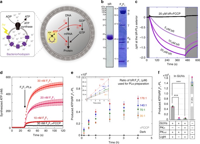
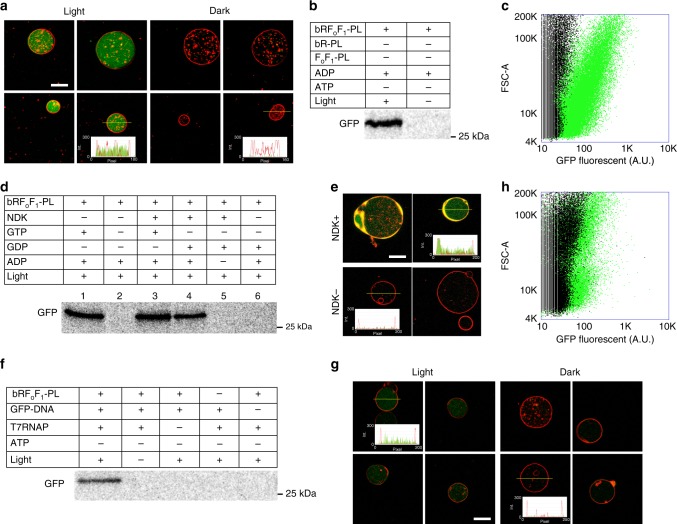
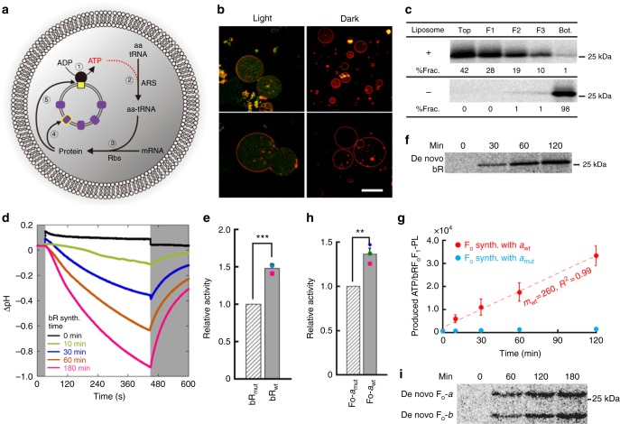
Attempts to construct an artificial cell have widened our understanding of living organisms. Many intracellular systems have been reconstructed by assembling molecules, however the mechanism to synthesize its own constituents by self-sufficient energy has to the best of our knowledge not been developed. Here, we combine a cell-free protein synthesis system and small proteoliposomes, which consist of purified ATP synthase and bacteriorhodopsin, inside a giant unilamellar vesicle to synthesize protein by the production of ATP by light. The photo-synthesized ATP is consumed as a substrate for transcription and as an energy for translation, eventually driving the synthesis of bacteriorhodopsin or constituent proteins of ATP synthase, the original essential components of the proteoliposome. The de novo photosynthesized bacteriorhodopsin and the parts of ATP synthase integrate into the artificial photosynthetic organelle and enhance its ATP photosynthetic activity through the positive feedback of the products. Our artificial photosynthetic cell system paves the way to construct an energetically independent artificial cell.


Conflict of interest statement

The authors declare no competing interests.

Figures

Fig. 1
Fig. 1
Light-driven adenosine triphosphate (ATP) synthesis by artificial organelle. a Schematics of the artificial photosynthetic cell encapsulating artificial organelle, which consists of bacteriorhodopsin (bR) and FoF1-ATP synthase (FoF1). Synthesized ATP are consumed as substrates for messenger RNA (mRNA) (➀), as energy for phosphorylation of guanosine diphosphate (GDP) (➁) or as energy for aminoacylation of transfer RNA (tRNA) (➂). b Sodium dodecyl sulfate-polyacrylamide gel electrophoresis (SDS-PAGE) analysis of purified bR and FoF1. The positions of molecular markers and FoF1 component proteins are indicated beside the gels. c Light-driven proton-pump activity of bR reconstituted in a proteoliposome (PL). Proton-pump activity of bR was measured by monitoring the proton concentration at the outside of bR-PLs where fluorescent proton-sensor ACMA (9-amino-6-chloro-2-methoxy acridine) was added. We defined as ΔpH = pH (original, outside) − pH (after illumination, outside). The ΔpH caused by bR activity was measured with the various bR concentrations as indicated. White and gray areas indicate light ON and OFF, respectively. An uncoupler, FCCP (carbonyl cyanide 4-(trifluoromethoxy) phenylhydrazone), was used as a control experiment. d ATP synthesis activity of FoF1 reconstituted as FoF1-PLs. ATP synthesis reactions were initiated by adding FoF1-PLs at 30 s with various FoF1 concentrations, as indicated. The synthesized ATP was measured by means of luciferin and luciferase (see Methods section for the experiment details). FCCP was used for control. e Light-driven ATP synthesis. The amount of the photosynthesized ATP by bRFoF1-PLs, which was constituted in various proportions of bR against FoF1, were measured by luciferin and luciferase. FCCP and dark conditions were also performed as controls. The inset indicates initial rate of the each PL. f Light-driven ATP synthesis inside giant unilamellar vesicle (GUV). bRFoF1-PLs were illuminated inside GUVs in the presence or absence of proteinase K (PK) that degrades the FoF1. The in vitro experiment was also performed for comparison. ***p < 0.001. P values were from two-sided t-test. All experiments were repeated at least three times, and their mean values and standard deviations (S.D.) are shown. Source data are provided as a Source Data file
Fig. 2
Fig. 2
Protein synthesis inside giant unilamellar vesicle (GUV) driven by light. Green fluorescent protein (GFP) was synthesized from its messenger RNA (mRNA) (ae) or DNA (fh) inside light illuminated GUV (a, c, eh) or in vitro (b, d). GFP was synthesized inside GUV (a) or in vitro (the PURE system) (b) in which the photosynthesized adenosine triphosphate (ATP) was consumed for the aminoacylation of transfer RNA (tRNA). The insets in a, e and g indicate plot profile of green and red colors on the thin yellow line. c Flow-cytometric analysis of the GUVs of a. The illuminated GUVs are shown as green, whereas the GUVs incubated in the dark are shown as black. The X- and Y-axes represent the fluorescent intensity and the area of forward scattering, respectively. d GFP synthesis coupled with guanosine 5’-triphosphate (GTP) generation. GFP was synthesized in the PURE system with or without nucleoside-diphosphate kinase (NDK), GTP, guanosine diphosphate (GDP) and adenosine 5’-diphosphate (ADP). e The same reactions as in lanes 4 and 6 of d were performed inside GUVs as indicated as NDK+ and NDK−, respectively. f GFP synthesis from its DNA. A gene of whole GFP was introduced in the PURE system with or without bRFoF1-PLs, T7 RNA polymerase (T7RNAP), ATP and light. g A small part of GFP (GFP11: 15 amino acids) was synthesized from its encoding DNA inside GUVs containing T7RNAP, another large part of GFP (GFP1-10) purified form E. coli cells, and the PURE system lacking NDK. h The same GUVs of g were analyzed by flow-cytometer as in d. The synthesized GFP in b, d, and f were labeled with [35S]methionine. Scale bar: 10 µm. Source data are provided as a Source Data file
Fig. 3
Fig. 3
Self-constituting protein synthesis in artificial photosynthetic cells. a Schematics of self-constituting protein synthesis. The numbers indicate the order of reactions; ➀: adenosine triphosphate (ATP) synthesis, ➁: aminoacylation of transfer RNA (tRNA) by aminoacyl-tRNA synthetase (ARS), ➂: translation by ribosomes (Rbs), ➃: de novo bacteriorhodopsin (bR) synthesis, and ➄: de novo Fo synthesis. b Light-induced bR-GFP synthesis in giant unilamellar vesicles (GUVs). Bar: 10 µm. c Membrane localization of bR. The bRs synthesized in the PURE system with or without liposomes were fractionated by ultra-centrifugation with sucrose cushion. The percentages of bR in each fraction (%Frac.) are indicated at the bottom of the gels. d Proton-pump activity of bR synthesized in the PURE system. The measurement was performed as in Fig. 1c. The reaction times of protein synthesis are indicated by different colors. The white and gray areas represent light ON and OFF, respectively. e Enhanced artificial organelle by de novo bR. Wild-type (bRwt) or mutant (bRmut) bRs were photosynthesized in the PURE system containing bRFoF1-PLs. The ATP concentrations at the time 2 h was measured and converted into ATP per proteoliposome (PL). The value of the de novo bRwt-containing PL was normalized to that of the de novo bRmut-containing PL. ***P < 0.001. f Sodium dodecyl sulfate–polyacrylamide gel electrophoresis (SDS-PAGE) analysis of the de novo photosynthesized bR. g Light-driven ATP synthesis by PLs consist of cell-free synthesized Fo. Wild-type (awt) or mutant (amut) a-subunit protein was synthesized together with b- and c-subunits in the PURE system containing purified F1 and bR-PLs. The measured ATP concentrations were converted into the produced ATP per PL. h Enhanced artificial organelle by de novo Fo. awt or amut was photosynthesized together with b- and c-subunits in the presence of purified F1 and bRFoF1-PLs. The ATP concentrations at the time 3 h was measured and converted into ATP per PL. The value of the de novo awt-containing PL was normalized by that of the de novo amut-containing PL. **P < 0.01. i SDS-PAGE analysis of the de novo photosynthesized Fo. P values were from two-side t-test. All experiments were performed at least three times and their means and S.D. are shown. Source data are provided as a Source Data file

References

    1. Rasmussen S, et al. Evolution. Transitions from nonliving to living matter. Science. 2004;303:963–965. doi: 10.1126/science.1093669. - DOI - PubMed
    1. Luisi, P. L. The Emergence of Life: From Chemical Origins to Synthetic Biology (Cambridge University Press, Cambridge, 2006).
    1. Mann S. Systems of creation: the emergence of life from nonliving matter. ACC Chem. Res. 2012;45:2131–2141. doi: 10.1021/ar200281t. - DOI - PubMed
    1. Hutchison CA, 3rd, et al. Design and synthesis of a minimal bacterial genome. Science. 2016;351:aad6253. doi: 10.1126/science.aad6253. - DOI - PubMed
    1. Xu C, Hu S, Chen X. Artificial cells: from basic science to applications. Mater. Today (Kidlington, Engl.) 2016;19:516–532. doi: 10.1016/j.mattod.2016.02.020. - DOI - PMC - PubMed

Publication types

MeSH terms

Substances