. 2019 Mar 22;9(1):5018.
doi: 10.1038/s41598-019-41123-2.

Etiology of 305 cases of refractory hematospermia and therapeutic options by emerging endoscopic technology

Affiliations

Etiology of 305 cases of refractory hematospermia and therapeutic options by emerging endoscopic technology

Liang-Gong Liao et al. Sci Rep. .

Abstract

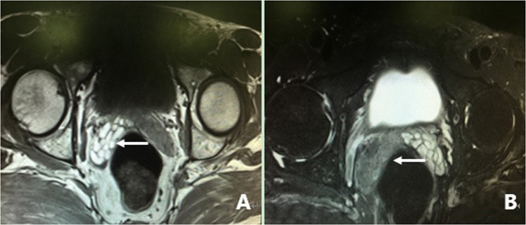
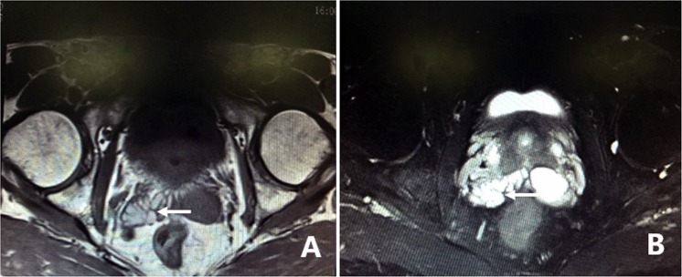
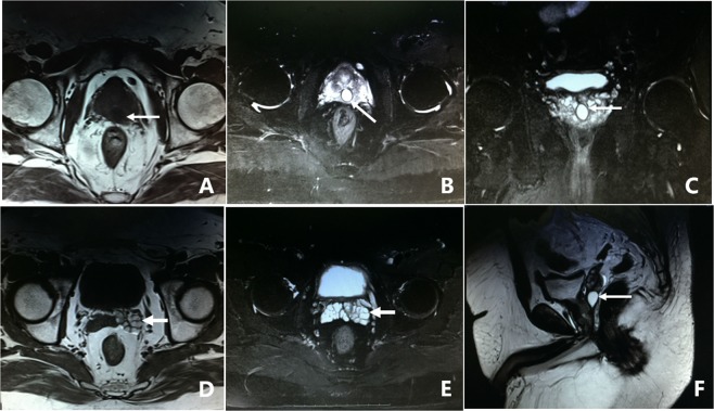
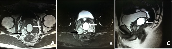
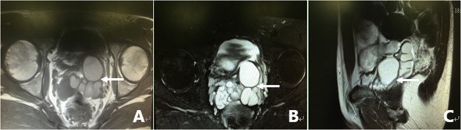
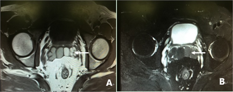
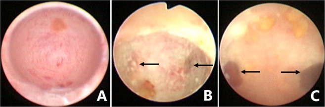
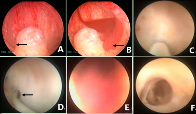
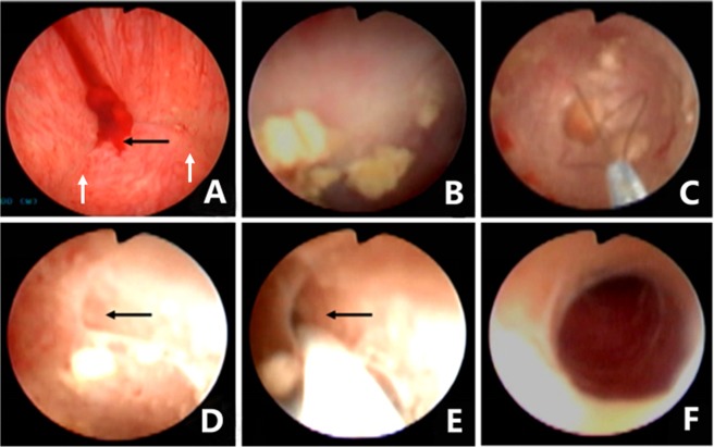
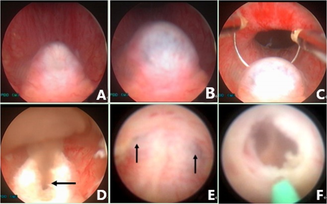
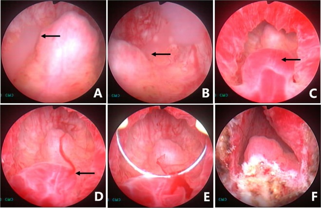
To investigate the surgical outcomes of vesiculoscopy on refractory hematospermia and ejaculatory duct obstruction (EDO), the clinical data (including pelvic magnetic resonance imaging (MRI) examinations and the long-term effects of endoscopic treatment) from 305 patients were analyzed. Four main etiologic groups were found on MRI. We found that 62.0% (189/305) of patients showed abnormal signal intensity in MRI investigations in the seminal vesicle (SV) area. Cystic lesions were observed in 36.7% (112/305) of the patients. The third sign was dilatation or enlargement of unilateral or bilateral SV, which were observed in 32.1% (98/305) of the patients. The fourth sign was stone formation in SV or in an adjacent cyst, which was present in 8.5% (26/305) of the patients. The transurethral endoscopy or seminal vesiculoscopy and the related procedures, including fenestration in prostatic utricle (PU), irrigation, lithotripsy, stone removal, biopsy, electroexcision, fulguration, or transurethral resection/incision of the ejaculatory duct (TURED/TUIED), chosen according to the different situations of individual patients were successfully performed in 296 patients. Fenestrations in PU+ seminal vesiculoscopy were performed in 66.6% (197/296) of cases. Seminal vesiculoscopy via the pathological opening in PU was performed in 10.8% (32/296) of cases. TURED/TUIED + seminal vesiculoscopy was performed in 12.8% (38/296) of cases, and seminal vesiculoscopy by the natural orifices of the ejaculatory duct (ED) was performed in 2.4% (7/296) of cases. Electroexcision and fulguration to the abnormal blood vessels or cavernous hemangioma at posterior urethra were performed in 7.4% (22/296) of cases. Two hundred and seventy-one patients were followed up for 6-72 months. The hematospermia of all the patients disappeared within 2-6 weeks, and 93.0% of the patients showed no further hematospermia during follow-up. No obvious postoperative complications were observed. The transurethral seminal vesiculoscopy technique and related procedures are safe and effective approaches for refractory hematospermia and EDO.


Conflict of interest statement

The authors declare no competing interests.

Figures

Figure 1
Figure 1
MRI image of a fresh hemorrhage in the right side of the SV. (A) This image shows high signal intensity in the right SV and low signal intensity in the left SV on T1WI. (B) This image shows low signal intensity in the right SV and high signal intensity in the left SV on T2WI. This pattern suggests that a fresh hemorrhage is present in the right SV.
Figure 2
Figure 2
MRI image of an old hemorrhage in the right side of the SV. (A) This image shows high signal intensity in the right SV and low signal intensity in the left SV on T1WI. (B) This image shows similar high signal intensity in both sides of the SV on T2WI. This pattern suggests that an old hemorrhage is present in the right SV. There are slight dilatations and cystic changes in the right and left sides of the SV, respectively.
Figure 3
Figure 3
MRI image of PUC. (A) There is a 1.0 × 1.2 cm low-intensity signal cyst in the midline of the prostate on T1WI. (B,C,F) The cyst presents as high-intensity signal on T2WI. (D,E) The left SV presents as high signal intensity on both T1WI and T2WI, and its width has expanded to 2.1 cm. This pattern suggests that the cyst is a PUC and is accompanied by enlargement and an old hemorrhage in the left SV.
Figure 4
Figure 4
MRI image of MDC. (A) There is a 3.4 × 4.5 cm high-intensity signal cyst in the midline of the prostate, accompanied by dilation and high-intensity signals in the right SV on T1WI. (B) Both the cyst and the bilateral SV present as high-intensity signal on T2WI. (C) The cyst extended beyond the posterosuperior margin of the prostate gland on the sagittal plane. This pattern suggests that the cyst is MDC, accompanied by dilation and an old hemorrhage in the right SV.
Figure 5
Figure 5
MRI image of SVC. (A) This image shows dilatation or enlargement in the left SV and presentation as a cystic structure with multiple cavities, with middle to high signal intensity in the left SV on T1WI. (B) This cyst presented as high signal intensity on T2WI, which is similar to that of the right SV. (C) The left SV presented as an obvious dilated cystic structure on the sagittal plane. This pattern suggests that the patient had a left SVC. There is also obvious dilation in the right SV.
Figure 6
Figure 6
MRI image of EDO. (A) The image shows obvious dilatation or enlargement in both sides of the SV. The SV presents intermediate-high signal intensity on T1WI. (B) Both sides of the SV present as low signal intensity on T2WI. This pattern suggests that EDO is accompanied with a fresh hemorrhage in both sides of the SV.
Figure 7
Figure 7
Different patterns under endoscopic observation in the PU. (A) The normal oval inner morphology of PU in some patients. (B) A pair of symmetrical translucent membranous weak areas was found at the 4 and 8 o’clock positions on the posterolateral wall of the PU in some patients. A small stone was also observed in the PU. (C) Bilateral ectopic passages of the EDs were observed at the 4 and 8 o’clock positions of the posterolateral wall of the PU in pathological conditions. Some small stones were observed in the PU.
Figure 8
Figure 8
Seminal vesiculoscopy and treatment by fenestration in the PU. (A) Endoscopic observation confirmed that the hemorrhage was not located on the right SV because normal jelly-like seminal fluid poured from the right ED orifice upon seminal vesicle massage. (B) Bloody fluid poured from the left ED orifice upon seminal vesicle massage, which indicated that the hemorrhage was located in the left SV. (C) A translucent membranous weak area was identified at the 4 o’clock position on the posterolateral wall of the utricle. (D) The soft tip of a guidewire was inserted along the channel without resistance for 3–5 cm, indicating that the tip had been inserted smoothly into the left SV. (E) The seminal vesiculoscope was inserted into the SV under the guidance of a guidewire. The SV was filled with a large quantity of red seminal fluid. (F) After irrigation and observation of the SV, the seminal vesiculoscope was withdrawn from the SV upon the formation of an ED short opening.
Figure 9
Figure 9
Seminal vesiculoscopy and treatment by the pathological opening in PU. (A) Bloody fluid poured from the orifice of the PU upon bilateral seminal vesicle massage, but nothing came from either of the ED orifices (white arrow). (B) The seminal vesiculoscope was inserted into the PU, and several stones were found in the PU. (C) The stones were removed by grasping forceps. (D) There are pathological openings in the PU at the 4 and 8 o’clock area in the PU (showing only the 4 o’clock opening). (E) The guidewire can be directly inserted into the SV through the opening. (F) The seminal vesiculoscope was inserted into the SV.
Figure 10
Figure 10
Seminal vesiculoscopy and treatment based on TURED. (A) Nothing came from either the ED orifice or the orifice of the PU upon bilateral SV massage, indicating the patients had complete EDO. (B) Transrectal massage to the medial area of the prostate showed that the PU opening is obstructed, and the verumontanum presented as a cystic mass with fluctuation. (C) Transurethral deroofing resection of the verumontanum was performed. (D) After transurethral deroofed resection of the verumontanum, chocolate-like fluid was observed emerging from the orifice of the PU. (E) Translucent membranous weak areas were identified at the 4 and 8 o’clock positions on the posterolateral wall of the utricle. (F) A holmium laser was used for fenestration.
Figure 11
Figure 11
Management of a cavernous hemangioma at the posterior urethra. (A) Normal white jelly-like seminal fluid poured from the right ED orifice upon seminal vesicle massage. (B) Normal white jelly-like seminal fluid poured from the left ED orifice upon seminal vesicle massage. (C) An obvious cavernous hemangioma with a size of about 5 × 5 mm was seen at about 5 mm away from the distal margin of the verumontanum. (D) Active bleeding was observed on the cavernous hemangioma. (E) The cavernous hemangioma was resected and/or fulgurated with an electric cutting loop. (F) The verumontanum area after the hemangioma was resected and/or fulgurated.

References

    1. Mulhall JP, Albertsen PC. Hemospermia: diagnosis and management. Urology. 1995;46:463–467. doi: 10.1016/S0090-4295(99)80256-8. - DOI - PubMed
    1. Han M, et al. Association of hemospermia with prostate cancer. J Urol. 2004;172:2189–2192. doi: 10.1097/01.ju.0000144565.76243.b1. - DOI - PubMed
    1. Polito M, et al. Hematospermia: diagnosis and treatment. Arch Ital Urol Androl. 2006;78:82–85. - PubMed
    1. Suh Y, et al. Etiologic classification, evaluation, and management of hematospermia. Transl Androl Urol. 2017;6:959–972. doi: 10.21037/tau.2017.06.01. - DOI - PMC - PubMed
    1. Li BJ, et al. Clinical analysis of the characterization of magnetic resonance imaging in 102 cases of refractory haematospermia. Andrology. 2013;1:948–956. doi: 10.1111/j.2047-2927.2013.00132.x. - DOI - PubMed

MeSH terms