Review
. 2019 Jun;26(6):981-993.
doi: 10.1038/s41418-019-0317-6. Epub 2019 Mar 22.

Decoding the sweet regulation of apoptosis: the role of glycosylation and galectins in apoptotic signaling pathways

Affiliations
Review

Decoding the sweet regulation of apoptosis: the role of glycosylation and galectins in apoptotic signaling pathways

Kamil Seyrek et al. Cell Death Differ. 2019 Jun.

Abstract

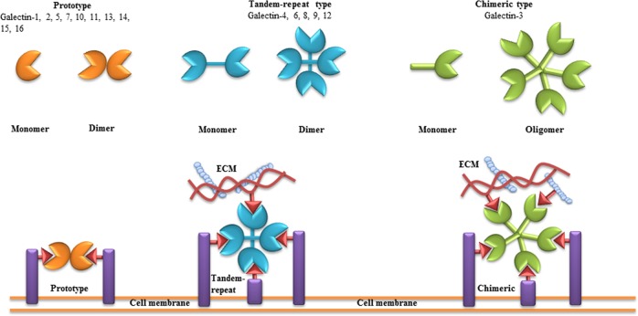
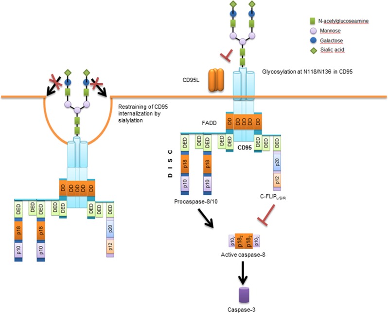
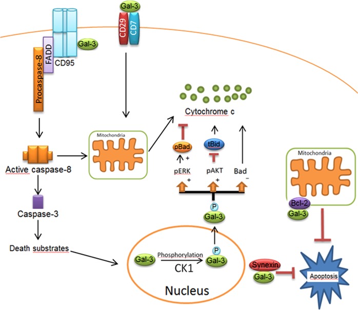
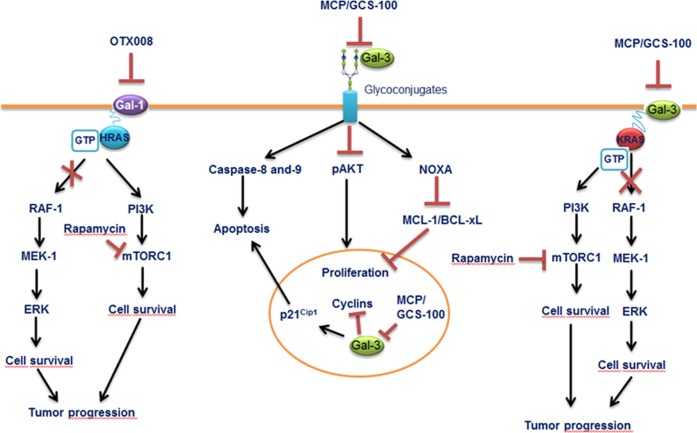
Glycosylation and glycan-binding proteins such as galectins play an important role in the control of cell death signaling. Strikingly, very little attention has been given so far to the understanding of the molecular details behind this key regulatory network. Glycans attached to the death receptors such as CD95 and TRAIL-Rs, either alone or in a complex with galectins, might promote or inhibit apoptotic signals. However, we have just started to decode the functions of galectins in the modulation of extrinsic and intrinsic apoptosis. In this work, we have discussed the current understanding of the glycosylation-galectin regulatory network in CD95- as well as TRAIL-R-induced apoptosis and therapeutic strategies based on targeting galectins in cancer.


Conflict of interest statement

The authors declare no competing interests.

Figures

Fig. 1
Fig. 1. Classification of galectins.
According to the number and arrangement of the carbohydrate recognition domains (CRDs), galectin family members are classified into three main types: proto, chimera, and tandem-repeat. Some galectins can self-associate into dimers or oligomers. The consensus sequence, which corresponds to the CRD, consists of ∼130 amino acids. Via their CRDs, galectins can interact with poly-N-acetyllactosamine (Gal-β(1-4)-GlcNAc, LacNAc)-based carbohydrates present in proteins, lipids, or other molecules. “One-CRD” galectins usually exist as dimers, whereas Gal-3 forms upon binding to Gal-β(1-4)-GlcNAc structures on cell surface and extracellular matrix. Tandem-repeat-type galectins comprise two homologous CRDs, separated by a short linker of up to 70 amino acids. Figure is modified from [100]
Fig. 2
Fig. 2. Glycosylation network of CD95 and CD95-mediated apoptosis.
Triggering CD95 by CD95L or agonistic antibodies induces its oligomerization and the subsequent formation of the death-inducing signaling complex (DISC). At the DISC, procaspase-8 undergoes dimerization in the death effector domain filaments that is followed by the activation of caspase-8. The mature caspase-8, consisting of two p18 and two p10 subunits, cleaves and activates downstream effector caspases, including caspase-3. CD95 is N-glycosylated in the extracellular domain, which can modulate the function of CD95 by several mechanisms discussed in the text
Fig. 3
Fig. 3. Apoptotic network of Gal-3 in extrinsic and intrinsic pathways.
Gal-3 comprises a serine phosphorylation site, which is involved in its translocation into the cytosol. Following the phosphorylation of Gal-3 by Casein Kinase 1 (CK1), Gal-3 translocates from the nucleus to the cytoplasm. The phosphorylated form of Gal-3(pGal-3) in the cytoplasm induces Bad phosphorylation via Erk pathway. pGal-3 promotes Akt activation, which blocks cleavage of Bid into tBid preventing mitochondrial outer membrane polarization. Gal-3 also attenuates cytochrome C release from the mitochondria by decreasing Bad expression. Synexin is required for Gal-3 translocation from the nucleus into the cytoplasm. In Type I cells, Gal-3 has been reported to interact with CD95, which results in high amounts of death-inducing signaling complex and active caspase-8. Extracellular Gal-3 binds to the CD29/CD7 complex, which triggers the activation of an intracellular apoptotic signaling cascade followed by cytochrome C release from mitochondria. Figure is modified from [32]
Fig. 4
Fig. 4. Mechanisms of action of galectin inhibitors.
Inhibition of Gal-1 by OTX008 results in HRAS mistargeting to the cell membrane, leading to the blockage of mitogen-activated protein kinase mitogenic pathway, whereas rapamycin decreases mammalian target of rapamycinsurvival signaling. Combination of OTX008 and rapamycin yields more effective treatment against tumor progression. Blockage of Gal-3 with MCP/GSC-100 activates caspase-8 and -9 pathways and induces NOXA protein, leading to decrease in MCL-1 and BCL-XL. Inhibition of nuclear Gal-3 induces cell-cycle inhibitor p21Cip1 expression and blocks the expression of cyclins, leading to cell death. Inhibition of Gal-3 with MCP/GSC-100 reduces KRAS-activated tumor progression. The combined treatment using MCP/GCS-100 and rapamycin together, however, has revealed more promising results in the KRAS mutant tumor progression experimental models. Figure is modified from [101]

References

    1. Elmore S. Apoptosis: a review of programmed cell death. Toxicol Pathol. 2007;35:495–516. doi: 10.1080/01926230701320337. - DOI - PMC - PubMed
    1. Igney FH, Krammer PH. Death and anti-death: tumour resistance to apoptosis. Nat Rev Cancer. 2002;2:277–88. doi: 10.1038/nrc776. - DOI - PubMed
    1. Weber CH, Vincenz C. The death domain superfamily: a tale of two interfaces? Trends Biochem Sci. 2001;26:475–81. doi: 10.1016/S0968-0004(01)01905-3. - DOI - PubMed
    1. Lavrik I, Golks A, Krammer PH. Death receptor signaling. J Cell Sci. 2005;118(Pt 2):265–7. doi: 10.1242/jcs.01610. - DOI - PubMed
    1. Krammer PH, Arnold R, Lavrik IN. Life and death in peripheral T cells. Nat Rev Immunol. 2007;7:532–42. doi: 10.1038/nri2115. - DOI - PubMed

Publication types