Myocardial strain imaging: review of general principles, validation, and sources of discrepancies
- PMID: 30903139
- PMCID: PMC6529912
- DOI: 10.1093/ehjci/jez041
Myocardial strain imaging: review of general principles, validation, and sources of discrepancies
Abstract
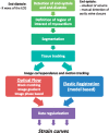
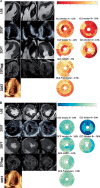
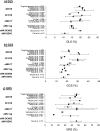
Myocardial tissue tracking imaging techniques have been developed for a more accurate evaluation of myocardial deformation (i.e. strain), with the potential to overcome the limitations of ejection fraction (EF) and to contribute, incremental to EF, to the diagnosis and prognosis in cardiac diseases. While most of the deformation imaging techniques are based on the similar principles of detecting and tracking specific patterns within an image, there are intra- and inter-imaging modality inconsistencies limiting the wide clinical applicability of strain. In this review, we aimed to describe the particularities of the echocardiographic and cardiac magnetic resonance deformation techniques, in order to understand the discrepancies in strain measurement, focusing on the potential sources of variation: related to the software used to analyse the data, to the different physics of image acquisition and the different principles of 2D vs. 3D approaches. As strain measurements are not interchangeable, it is highly desirable to work with validated strain assessment tools, in order to derive information from evidence-based data. There is, however, a lack of solid validation of the current tissue tracking techniques, as only a few of the commercial deformation imaging softwares have been properly investigated. We have, therefore, addressed in this review the neglected issue of suboptimal validation of tissue tracking techniques, in order to advocate for this matter.
Keywords: cMR; echocardiography; feature tracking; review; speckle tracking imaging; strain; tagging.
© The Author(s) 2019. Published by Oxford University Press on behalf of the European Society of Cardiology.
Figures


References
-
- Mirsky I, Parmley WW.. Assessment of passive elastic stiffness for isolated heart muscle and the intact heart. Circ Res 1973;33:233–43. - PubMed
-
- Domanski MJ, Follmann D, Mirsky II.. A new approach to assessing regional and global myocardial contractility. Echocardiography 1997;14:1–8. - PubMed
-
- Zerhouni EA, Parish DM, Rogers WJ, Yang A, Shapiro EP.. Human heart: tagging with MR imaging–a method for noninvasive assessment of myocardial motion. Radiology 1988;169:59–63. - PubMed
-
- Sutherland GR, Stewart MJ, Groundstroem KW, Moran CM, Fleming A, Guell-Peris FJ.. Color Doppler myocardial imaging: a new technique for the assessment of myocardial function. J Am Soc Echocardiogr 1994;7:441–58. - PubMed
Publication types
MeSH terms
LinkOut - more resources
Full Text Sources
Other Literature Sources
Medical
Research Materials

