. 2019 Jun;145(6):1405-1416.
doi: 10.1007/s00432-019-02901-7. Epub 2019 Mar 22.

NOD1 and NOD2 of the innate immune system is differently expressed in human clear cell renal cell carcinoma, corresponding healthy renal tissue, its vasculature and primary isolated renal tubular epithelial cells

Affiliations

NOD1 and NOD2 of the innate immune system is differently expressed in human clear cell renal cell carcinoma, corresponding healthy renal tissue, its vasculature and primary isolated renal tubular epithelial cells

Lilli Mey et al. J Cancer Res Clin Oncol. 2019 Jun.

Abstract

Purpose: NOD1 and NOD2 (nucleotide-binding oligomerization domain)-receptors are intracellular receptors and belong to the family of pattern recognition receptors being present in both human and murine renal tubular cells. Besides, NOD1 has been proved to promote apoptosis, upon its overexpression. Hence, we aimed to investigate NOD1 and NOD2 expression in human clear cell renal cell carcinoma (ccRCC).

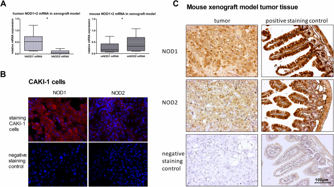
Methods: Tumor and corresponding adjacent healthy tissues from 41 patients with histopathological diagnosis of ccRCC as well as primary isolated renal tubular epithelial cells (TECs) and tumor tissue from a murine xenograft model using CAKI-1 ccRCC cells were analyzed.

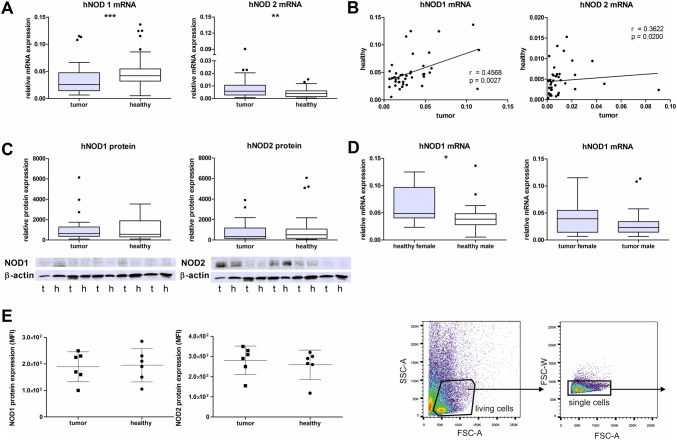
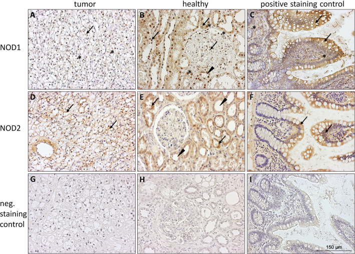
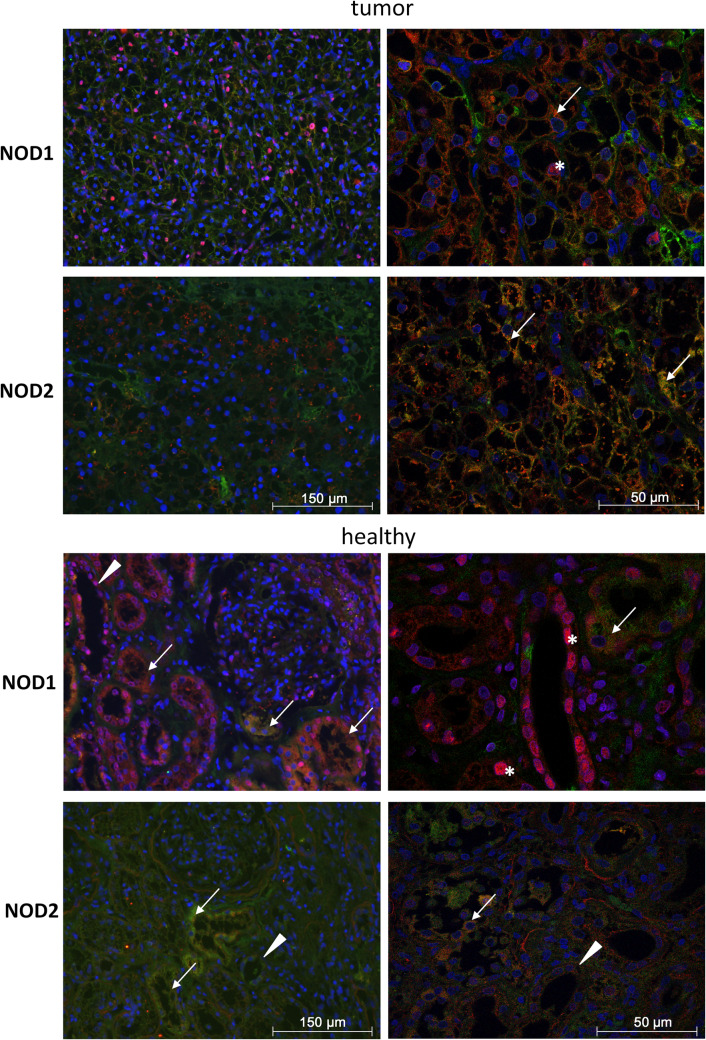
Results: NOD1 and NOD2 mRNA was constitutively expressed in both tumor and adjacent healthy renal tissue, with NOD1 being significantly lower and in contrast NOD2 significantly higher expressed in tumor tissue compared to healthy tissues. Immunohistochemically, NOD1 was located not only in the cytoplasm, but also in the nucleus in ccRCC tissue whereas NOD2 was solely localized in the cytoplasm in both human ccRCC as well as in the healthy tubular system. Focusing on the vasculature, NOD2 displayed broader expression than NOD1. In primary TECs as well as CAKI-1 cells NOD1 and NOD2 was constitutively expressed and increasable upon LPS stimulation. In the mouse xenograft model, human NOD1 mRNA was significantly higher expressed compared to NOD2. In contrast hereto, we observed a shift towards lower mouse NOD1 compared to NOD2 mRNA expression.

Conclusion: In view of reduced apoptosis-associated NOD1 expression in ccRCC tissue opposed to higher expression of NOD2 in tumor vasculature, inducibility of NOD expression in TECs as well as the detected shift of NOD1 and NOD2 expression in the mouse xenograft model, modulation of NOD receptors might, therefore, provide a molecular therapeutic approach in ccRCC.

Keywords: Clear cell renal cell carcinoma; Innate immunity; Kidney cancer; NLR.


Conflict of interest statement

The authors declare that they have no conflict of interest and that the results presented in this paper have not been published previously in whole or part.

Figures

Fig. 1
Fig. 1
mRNA and protein expression in ccRCC and healthy human kidney tissue. a NOD1 and NOD2 mRNA expression normalized to its RPL-PO content in tumor and adjacent healthy renal tissue (n = 33) b and their correlation (n = 33) (Spearman’s rank correlation test). d NOD1 mRNA in female and male subjects. c Protein expression of NOD1 (110 kDa), NOD2 (110 kDa) and β-actin (43 kDa) (n = 23) with representative western blots. e Mean fluorescence intensity in flow cytometric analysis of intracellular NOD1 and NOD2 (n = 6). NOD mRNA and protein expression are represented in box blots to depict the distributions of the relative expression values. *p < 0.5, **p < 0.01, ***p < 0.001
Fig. 2
Fig. 2
Immunohistochemical staining for NOD1 and NOD2 receptors in ccRCC and healthy human kidney tissue. a Localization of NOD1 in the cytoplasm (arrows) and nucleus (*) of ccRCC cells. b Arrows indicate localization of NOD1 receptor in the cytoplasm and nucleus (*) of the tubular system, podocytes and vascular cells in healthy kidney. Arrowhead indicates a tubule. c High expression of NOD1 receptor in the lamina epithelialis (arrows). Nuclear and perinuclear staining of cells of the lamina propria (*) in the mucosa of human ileum as positive control. d NOD2 localization in the cytoplasm (arrows) of ccRCC cells. e NOD2 is located in cytoplasm primarily in the tubules (arrows and arrowheads). f NOD2 localization (arrows) in the mucosa of human ileum as a positive control. g Negative staining control of ccRCC and healthy tissue has been performed in the absence of the primary antibodies. h Negative staining control of ccRCC and healthy tissue has been performed in the absence of the primary antibodies. i Negative staining control of human ileum has been performed in the absence of the primary antibodies. (200 fold magnification)
Fig. 3
Fig. 3
Immunofluorescence staining of NOD1 and NOD2 receptor in ccRCC and healthy tissue. Immunofluorescence staining of NOD1 and NOD2 receptor in red (Cy3) and proximal tubule specific CD13 in green (Alexa 488) in ccRCC and healthy renal tissue. Co-localization appear as merge in yellow. Nuclear staining was performed using DAPI. Assessment with confocal microscopy (right pictures). Nuclear (white*) and cytoplasmic (white arrows and arrowheads) localization of NOD1 and NOD2 receptor. Higher expression of NOD1 receptor in healthy kidney, especially in proximal (white arrows), distal (white arrowheads) and collecting duct. Co-localization of NOD1 with CD13 in the proximal tubuli (white arrows). High NOD2 expression occurs in tumor tissue (left pictures in 200-fold and right pictures in 600-fold magnification)
Fig. 4
Fig. 4
Immunohistochemical staining for NOD1 and NOD2 receptors in ccRCC and healthy tissue vasculature. Localization of NOD1 or NOD2 in endothelial cells (arrows) and tunica media (arrowhead). Negative staining control of ccRCC and healthy tissue has been performed in the absence of the primary antibodies. (200 fold magnification)
Fig. 5
Fig. 5
Immunofluorescence staining of NOD1 and NOD2 receptor in primary isolated ccRCC cells and corresponding renal tubular epithelial cells (TECs). NOD1 and NOD2 expression in red (Cy3) in primary isolated tumor cells and renal tubular epithelial cells (TECs) of healthy tissue. TECs were untreated or incubated with LPS (10 µg/ml) for 4 h. Nuclear DAPI staining. Negative staining control was performed without the use of primary antibody. (200-fold magnification)
Fig. 6
Fig. 6
NOD1 and NOD2 expression in a heterotopic tumor model in the mouse. a Human NOD1 and NOD2 mRNA as well as mouse NOD1 and NOD2 mRNA expression normalized to its 18 s content in a heterotopic tumor model in the mouse (n = 6) presented in box blots to depict the distributions of the relative expression values. *p < 0.5. b Immunofluorescence staining of NOD1 and NOD2 receptor in CAKI-1 cells. NOD1 and NOD2 expression in red (Cy3) in untreated CAKI-1 cells. Nuclear DAPI staining. Negative staining control was performed without the use of primary antibody (200-fold magnification). c Immunohistochemical staining of NOD1 and NOD2 receptor in tumor areas of the xenograft model and mouse ileum as positive control. Negative staining control was performed without the use of primary antibody (200-fold magnification)

Similar articles

Cited by

References

    1. Baer PC, Nockher WA, Haase W, Scherberich JE (1997) Isolation of proximal and distal tubule cells from human kidney by immunomagnetic separation. Kidney Int 52:1321–1331 - PubMed
    1. Bamias A et al (2017) Current clinical practice guidelines for the treatment of renal cell carcinoma: a systematic review and critical evaluation. Oncologist 22:667–679. 10.1634/theoncologist.2016-0435 - PMC - PubMed
    1. Bertin J et al (1999) Human CARD4 protein is a novel CED-4/Apaf-1 cell death family member that activates NF-kappaB. J Biol Chem 274:12955–12958 - PubMed
    1. Braunstein MJ, Kucharczyk J, Adams S (2018) Targeting toll-like receptors for cancer therapy. Target Oncol. 10.1007/s11523-018-0589-7 - PubMed
    1. Bray F, Ferlay J, Soerjomataram I, Siegel RL, Torre LA, Jemal A (2018) Global cancer statistics 2018: GLOBOCAN estimates of incidence and mortality worldwide for 36 cancers in 185 countries. Cancer J Clin. 10.3322/caac.21492 - PubMed

MeSH terms

Grants and funding

LinkOut - more resources