. 2019 May 2;74(3):481-493.e6.
doi: 10.1016/j.molcel.2019.02.017. Epub 2019 Mar 20.

Retapamulin-Assisted Ribosome Profiling Reveals the Alternative Bacterial Proteome

Affiliations

Retapamulin-Assisted Ribosome Profiling Reveals the Alternative Bacterial Proteome

Sezen Meydan et al. Mol Cell. .

Abstract

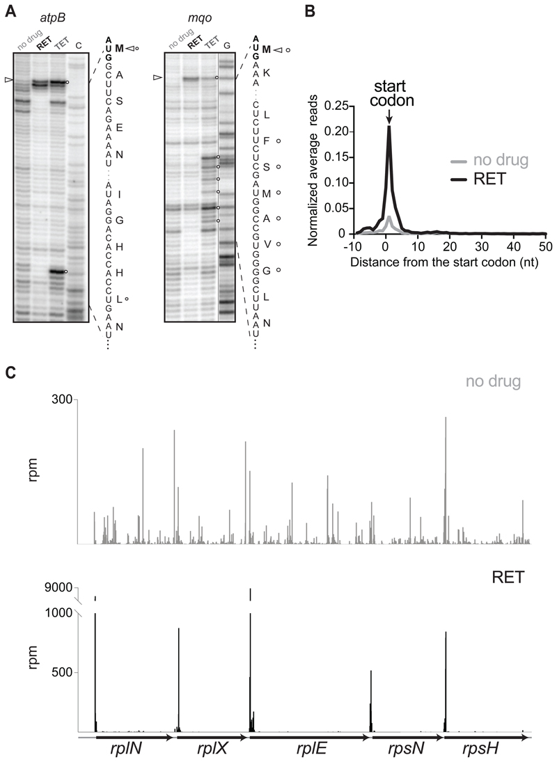
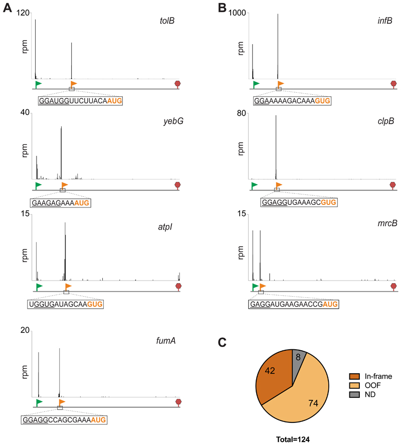
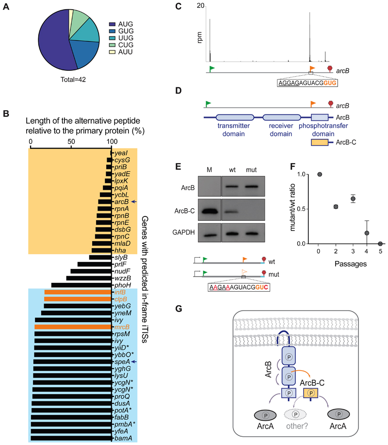
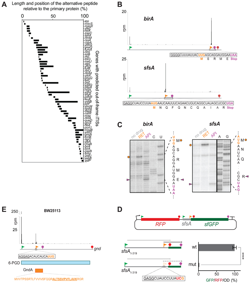
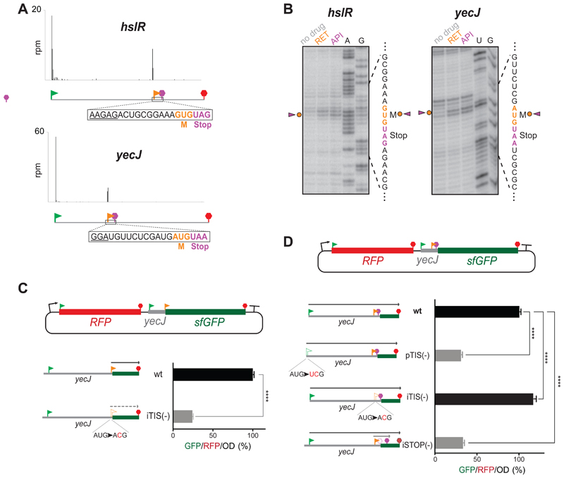
The use of alternative translation initiation sites enables production of more than one protein from a single gene, thereby expanding the cellular proteome. Although several such examples have been serendipitously found in bacteria, genome-wide mapping of alternative translation start sites has been unattainable. We found that the antibiotic retapamulin specifically arrests initiating ribosomes at start codons of the genes. Retapamulin-enhanced Ribo-seq analysis (Ribo-RET) not only allowed mapping of conventional initiation sites at the beginning of the genes, but strikingly, it also revealed putative internal start sites in a number of Escherichia coli genes. Experiments demonstrated that the internal start codons can be recognized by the ribosomes and direct translation initiation in vitro and in vivo. Proteins, whose synthesis is initiated at internal in-frame and out-of-frame start sites, can be functionally important and contribute to the "alternative" bacterial proteome. The internal start sites may also play regulatory roles in gene expression.

Keywords: alternative initiation; arcB; internal genes; retapamulin; ribosome profiling; rpn; speA; translation initiation.


Conflict of interest statement

Declarations of Interest

The authors declare no competing interests.

Figures

Figure 1
Figure 1. RET specifically arrests ribosomes at translation initiation sites
(A) Toeprinting analysis showing retapamulin (RET) (triangles)- and tetracycline (TET) (circles)- induced translation arrest sites during cell-free translation of two model E. coli genes. ‘C’ and ‘G’ indicate sequencing lanes. Nucleotide and encoded amino acid sequences are shown. (B) Metagene analysis plot representing normalized average relative density reads in the vicinity of the annotated start codons of the genes of E. coli cells treated or not with RET. (C) Ribosome footprints density within the spc operon in cells treated or not with RET. See also Figures S1.
Figure 2
Figure 2. Ribo-RET reveals the presence of iTISs in many bacterial genes
(A) Examples of Ribo-RET profiles of E. coli genes with newly detected iTISs. The annotated pTISs are marked with green flags and stop codons are shown with red stop signs; orange flags show the iTISs. iTIS start codons are highlighted in orange and the SD-like sequences are underlined. (B) Ribo-RET profiles of infB, clpB, mrcB, the three E. coli genes where iTISs had been previously characterized. (C) The iTISs common between the E. coli BW25113 and BL21 strains. ND (not determined): the internal Ribo-RET peaks not associated with the known start codons. See also Table S1.
Figure 3
Figure 3. In-frame internal initiation can generate functional N-terminally truncated proteins
(A) The frequency of various putative start codons at the in-frame iTISs. (B) The relative length of the predicted alternative proteins, products of internal initiation, in comparison with the main protein. The known examples of genes with in-frame iTISs (Figure 2B) are in orange. The genes with the iTISs located within the 3’ or 5’ quartile of the gene length are boxed in yellow or blue, respectively. Asterisks show genes with pTIS re-annotation proposed based on TET-assisted Ribo-seq, (Nakahigashi et al., 2016). Arrows point at arcB and speA genes further analyzed in this work. (C) Ribo-RET profile of arcB showing ribosomal density peaks at the pTIS (green flag) and the iTIS (orange flag). (D) Schematics of the functional domains of ArcB. The putative alternative ArcB-C protein would encompass the phosphotransfer domain. (E) Western blot analysis of the C-terminally 3XFLAG-tagged translation products of the arcB gene expressed from a plasmid in E. coli cells. Inactivation of iTIS by the indicated mutations (mut) abrogates production of ArcB-C. Lane M: individually-expressed marker protein ArcB-C-3XFLAG (see STAR Methods). (F) Functional iTIS in arcB facilitates growth under low oxygen conditions. BW25113 ΔarcB E. coli cells, expressing from a plasmid either wt arcB or mutant arcB with inactivated iTIS, were co-grown in low oxygen conditions and the ratio of mutant to wt cells was analyzed (see Start Methods for details). Error bars represent deviation from the mean (n=2). (G) The phosphorelay across the wt ArcB domains results in the activation of the response regulator ArcA (Alvarez et al., 2016). Diffusible ArcB-C could amplify the signal capabilities of the ArcBA system and/or enable cross-talk with other response regulators. See also Figure S2.
Figure 4
Figure 4. OOF iTISs revealed by Ribo-RET can direct initiation of translation
(A) The length and location of the alternative OOF protein-coding segments relative to the main ORF. (B) Ribo-RET profiles of birA and sfsA genes with putative OOF iTISs. The iTIS OOF start codon and the corresponding stop codon are indicated by orange flag and stop sign, respectively. The sequences of the alternative ORFs are shown. (C) Toeprinting analysis reveals that RET arrests ribosomes at the start codons (orange circles) of the alternative ORFs within the birA and sfsA genes; termination inhibitor Api137 arrests translation at the stop codons (purple arrowheads) of those ORFs. The nucleotide and amino acid sequences of the alternative ORFs are shown. Sequencing lanes are indicated. (D) In the cell, translation is initiated at the sfsA OOF iTIS. Schematics of the RFP/sfGFP reporter plasmid. The rfp and sf-gfp genes are co-transcribed. The sfGFP-coding sequence is expressed from the iTIS (orange flag) and is OOF relative to the sfsA pTIS start codon (green flag). The first stop codon in-frame with the pTIS (red) and the stop codon in-frame with the iTIS (orange) are indicated. The bar graph shows the change in relative green fluorescence in response to the iTIS start codon mutation (mut). The values represent the mean ± standard deviation from technical replicates (n=6). Two-tailed unpaired t-test. (E) Ribo-RET snapshots of the gnd gene revealing the putative location of the start codon of the alternative ORF. The amino acid sequence of the alternative ORF product GndA is shown in orange; the tryptic peptide identified by mass spectrometry (Yuan et al., 2018) is underlined. See also Figure S5 and Table S1.
Figure 5
Figure 5. Start-Stops within E. coli genes
(A) Representative Ribo-RET profiles revealing start-stops. The SD-like sequences are underlined. (B) Toeprinting analysis shows ribosomes stalled at the start codons of the start-stop sites in response to the presence of initiation (RET) and termination (API) inhibitors. The start and stop codons of the start-stop sites are indicated by orange and purple characters, respectively. (C) The start codon of the yecJ start-stop can direct initiation of translation in vivo. sfGFP expression in the RFP/sfGFP reporter is directed by the start codon of the yecJ start-stop (orange flag). The relative translation efficiency was estimated by measuring GFP/RFP/OD (%) ratio. The expression of sf-gfp is severely abrogated by a mutation that disrupts the start-stop initiation codon [iTIS (-)]. The values represent the standard deviation from the mean in technical replicates (n=3). Two-tailed unpaired t-test. (D) Start-stop impacts expression of the yecJ gene. The expression of the YecJGFP chimeric protein is controlled by yecJ pTIS (green flag) (gfp sequence is in 0-frame relative to pTIS). The reporter expression increases by ~16% when the start codon of the start-stop site is disrupted by a mutation [(iTIS(-)]. Mutating the stop codon of the start-stop site expands the length of the translated OOF coding sequence and results in severe inhibition of the main frame translation. The error bars represent standard deviation from the mean in technical triplicates (n=3). Two-tailed unpaired t-test. See also Figure S5 and Table S2.

References

    1. Alvarez AF, Barba-Ostria C, Silva-Jimenez H, Georgellis D. Organization and mode of action of two component system signaling circuits from the various kingdoms of life. Environ Microbiol. 2016;18:3210–3226. - PubMed
    1. Alvarez AF, Georgellis D. In vitro and in vivo analysis of the ArcB/A redox signaling pathway. Methods Enzymol. 2010;471:205–228. - PubMed
    1. Atkins JF, Loughran G, Bhatt PR, Firth AE, Baranov PV. Ribosomal frameshifting and transcriptional slippage: From genetic steganography and cryptography to adventitious use. Nucleic Acids Res. 2016;44:7007–7078. - PMC - PubMed
    1. Baba T, Ara T, Hasegawa M, Takai Y, Okumura Y, Baba M, Datsenko KA, Tomita M, Wanner BL, Mori H. Construction of Escherichia coli K-12 in-frame, single-gene knockout mutants: the Keio collection. Mol Syst Biol. 2006;2 2006 0008. - PMC - PubMed
    1. Baek J, Lee J, Yoon K, Lee H. Identification of unannotated small genes in Salmonella . G3 (Bethesda) 2017;7:983–989. - PMC - PubMed

Publication types

MeSH terms