

Unusual presentation of extramedullary haematopoiesis in a young boy
- PMID: 30904883
- PMCID: PMC6453391
- DOI: 10.1136/bcr-2018-227199
Unusual presentation of extramedullary haematopoiesis in a young boy
Abstract
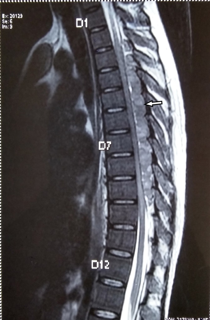
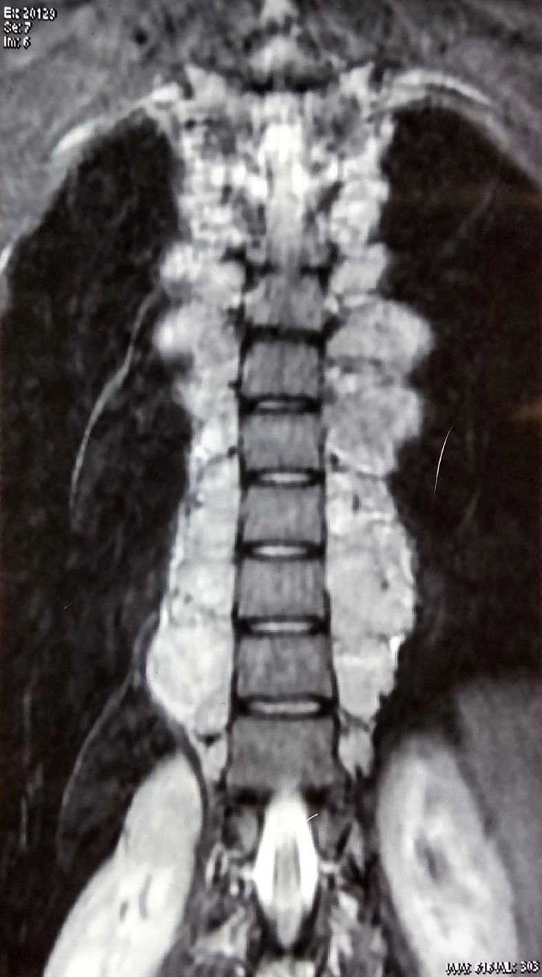
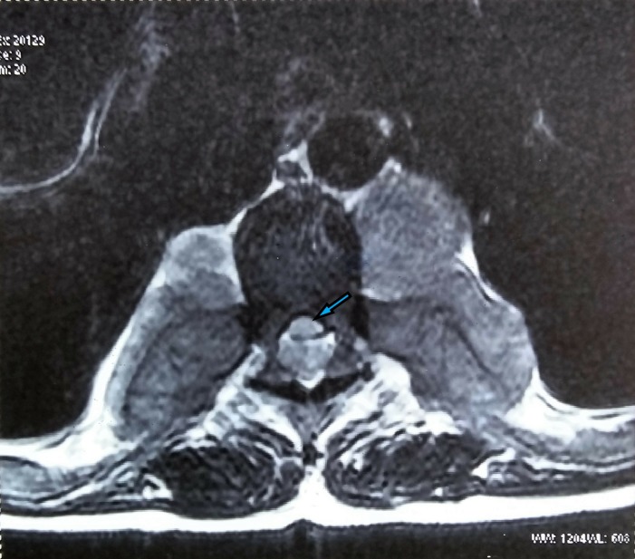
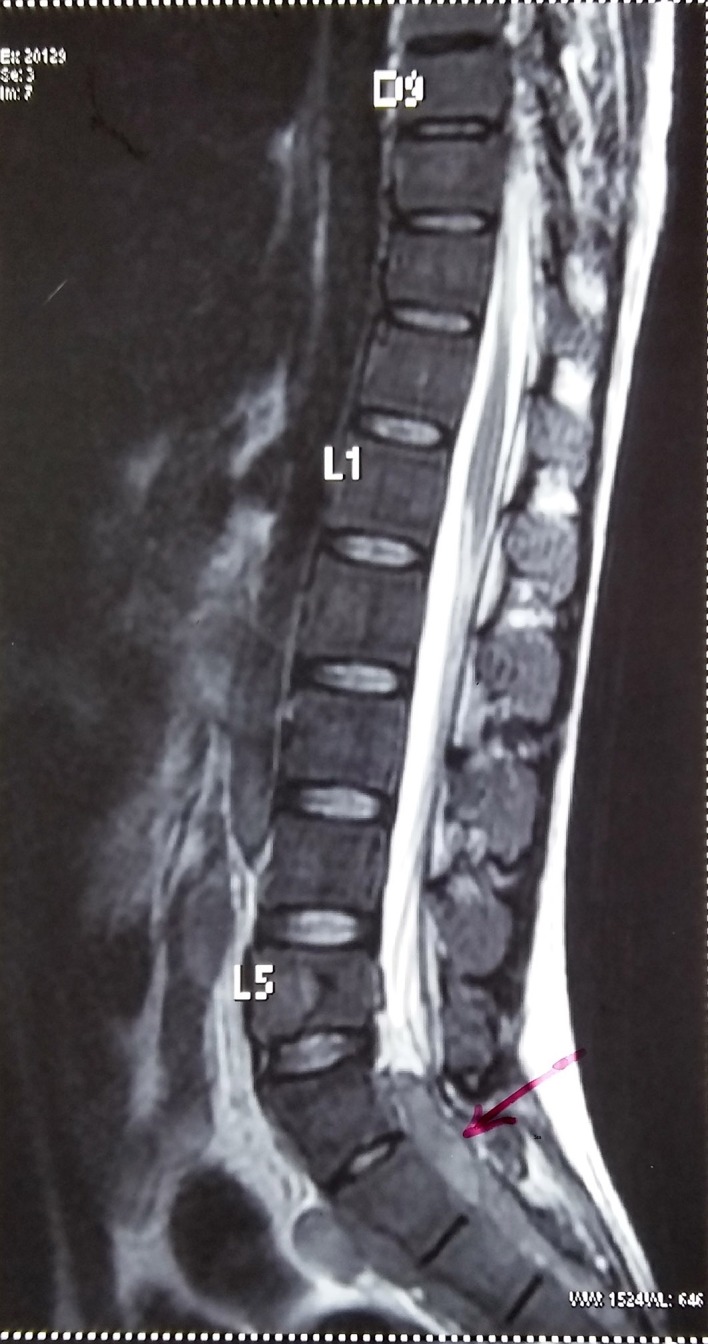
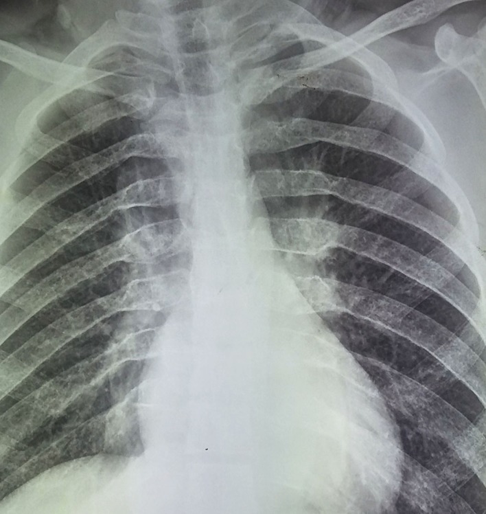
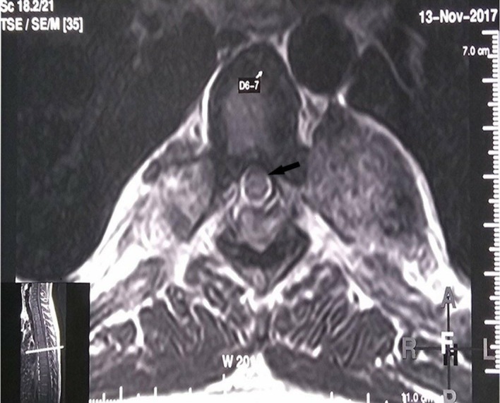
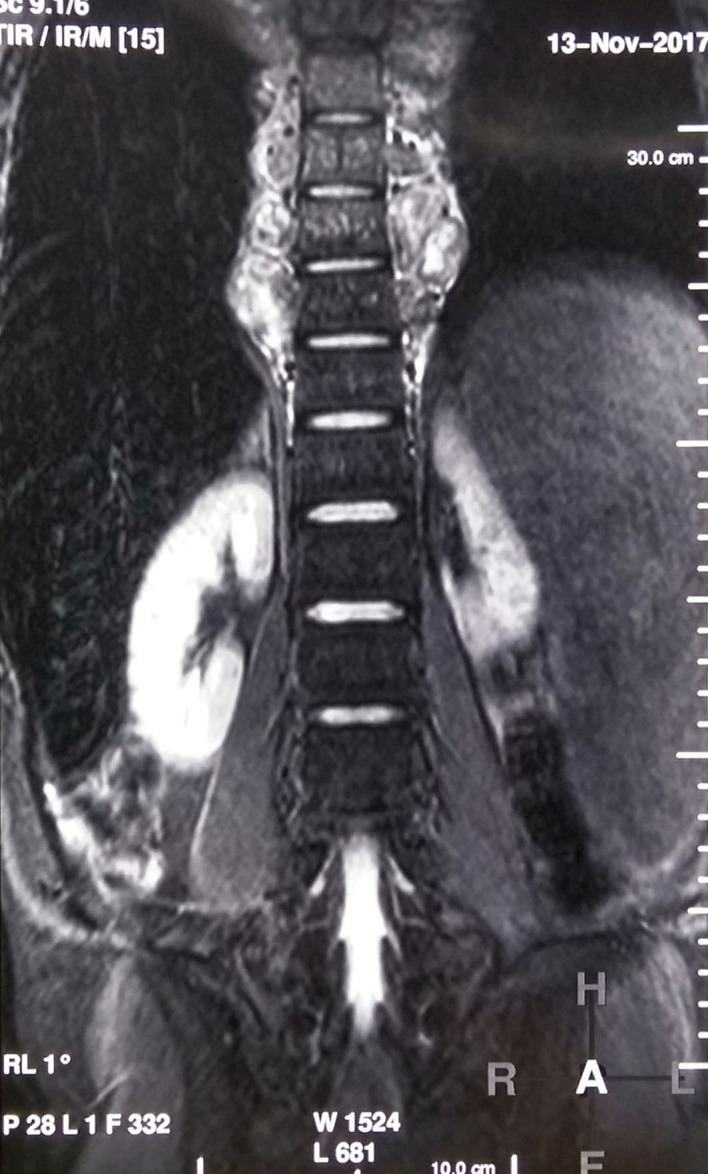
Acute transverse myelopathy in a young person may be due to infection, postinfective or inflammatory demyelination, or vascular causes. Rarely, a completely reversible cause of acute transverse myelopathy may be seen, as described here in our case of transverse myelopathy due to extramedullary haematopoiesis (EMH). An 18-year-old man who had a history of a lone blood transfusion at age of 7 years presented with paraplegia. MRI showed multiple epidural space masses of EMH compressing the spinal cord. He was detected to have thalassaemia intermedia and was treated with blood transfusions, steroids and radiotherapy to the involved paraspinal areas. He recovered fully over 15 days and remained symptom free at 6 months.
Keywords: haematology (incl blood transfusion); spinal cord.
© BMJ Publishing Group Limited 2019. No commercial re-use. See rights and permissions. Published by BMJ.
Conflict of interest statement
Competing interests: None declared.
Figures
References
-
- Amirjamshidi A, Abbassioun K, Ketabchi SE. Spinal extradural hematopoiesis in adolescents with thalassemia. Report of two cases and a review of the literature. Childs Nerv Syst 1991;7:223–5. - PubMed
-
- Weatherall DJ, Clegg JB. The thalassemia syndromes. 4th edn Oxford: Blackwell Scientific Publications, 2001.
-
- Gatto I, Terrana V, Biondi L. [Compression of the spinal cord due to proliferation of bone marrow in epidural space in a splenectomized person with Cooley’s disease]. Haematologica 1954;38:61–76. - PubMed
Publication types
MeSH terms
LinkOut - more resources
Full Text Sources