

Platinum sensitive carcinoma of ovary relapsed as pericardial effusion with cardiac tamponade
- PMID: 30904894
- PMCID: PMC6510153
- DOI: 10.1136/bcr-2018-228268
Platinum sensitive carcinoma of ovary relapsed as pericardial effusion with cardiac tamponade
Abstract
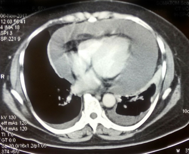
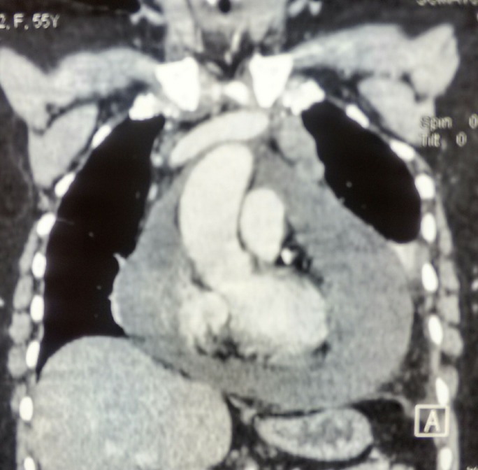
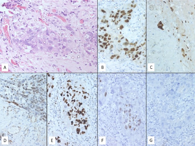
Epithelial ovarian cancers typically spread by intraperitoneal exfoliation and retroperitoneal lymph nodal involvement along the ovarian vascular supply. Pericardial involvement in ovarian malignancies is very rare with only few cases reported in the literature. Malignancy is the most common cause for pericardial effusion in the western world. In this case report, we present a 58-year-old woman treated for high-grade serous carcinoma of the ovary in 2010, relapsed with pericardial effusion and cardiac tamponade in 2017. Imaging studies revealed gross pericardial effusion. Two-dimensional echocardiogram showed massive pericardial effusion, with cardiac tamponade, New York Heart Association-IV. Pericardiocentesis and pigtail drain was placed under echo guidance. Immunocytochemistry has confirmed the tumour cells to be of the ovarian origin. The patient underwent surgical pericardial window via thoracotomy, followed by paclitaxel and carboplatin-based chemotherapy and olaparib maintenance.
Keywords: cancer - see oncology; chemotherapy; gynecological cancer.
© BMJ Publishing Group Limited 2019. No commercial re-use. See rights and permissions. Published by BMJ.
Conflict of interest statement
Competing interests: None declared.
Figures

References
-
- Blich M, Malkin L, Kapeliovich M. Malignant pericardial tamponade secondary to papillary serous adenocarcinoma of the ovary. Isr Med Assoc J 2007;9:337–8. - PubMed
Publication types
MeSH terms
Substances
LinkOut - more resources
Full Text Sources
Medical