. 2019 Jun;79(8):840-855.
doi: 10.1002/pros.23789. Epub 2019 Mar 24.

High levels of PIWI-interacting RNAs are present in the small RNA landscape of prostate epithelium from vitamin D clinical trial specimens

Affiliations

High levels of PIWI-interacting RNAs are present in the small RNA landscape of prostate epithelium from vitamin D clinical trial specimens

Bethany Baumann et al. Prostate. 2019 Jun.

Abstract

Background: Vitamin D, a hormone that acts through the nuclear vitamin D receptor (VDR), upregulates antitumorigenic microRNA in prostate epithelium. This may contribute to the lower levels of aggressive prostate cancer (PCa) observed in patients with high serum vitamin D. The small noncoding RNA (ncRNA) landscape includes many other RNA species that remain uncharacterized in prostate epithelium and their potential regulation by vitamin D is unknown.

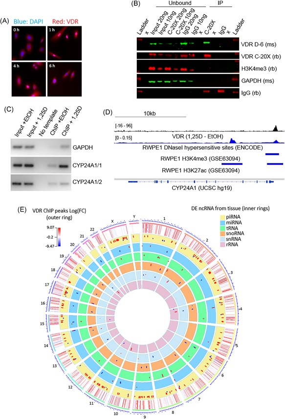
Methods: Laser capture microdissection (LCM) followed by small-RNA sequencing was used to identify ncRNAs in the prostate epithelium of tissues from a vitamin D-supplementation trial. VDR chromatin immunoprecipitation-sequencing was performed to identify vitamin D genomic targets in primary prostate epithelial cells.

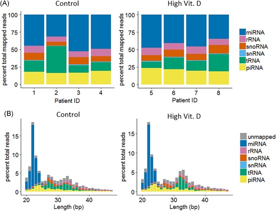
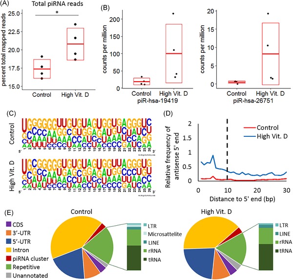
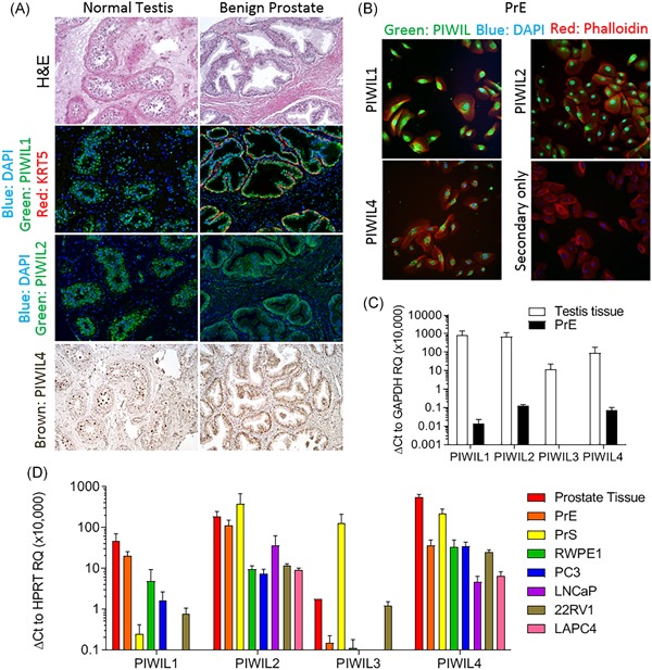
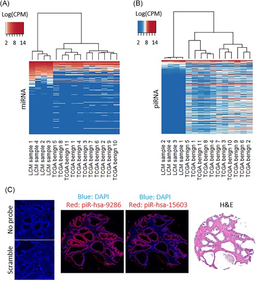
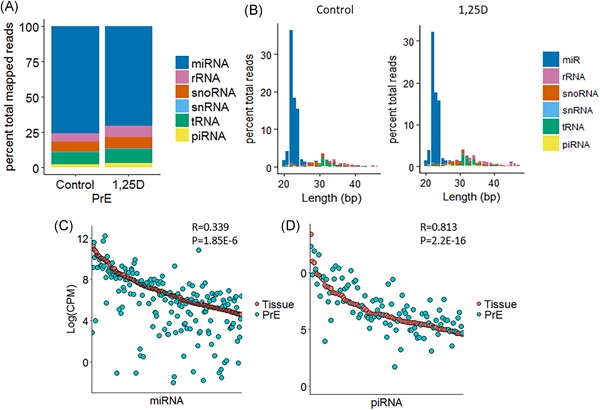
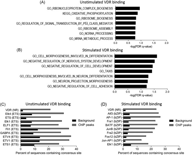
Results: Isolation of epithelium by LCM increased sample homogeneity and captured more diversity in ncRNA species compared with publicly available small-RNA sequencing data from benign whole prostate. An abundance of PIWI-interacting RNAs (piRNAs) was detected in normal prostate epithelium. The obligate binding partners of piRNAs, PIWI-like (PIWIL) proteins, were also detected in prostate epithelium. High prostatic vitamin D levels were associated with increased expression of piRNAs. VDR binding sites were located near several ncRNA biogenesis genes and genes regulating translation and differentiation.

Conclusions: Benign prostate epithelium expresses both piRNA and PIWIL proteins, suggesting that these small ncRNA may serve an unknown function in the prostate. Vitamin D may increase the expression of prostatic piRNAs. VDR binding sites in primary prostate epithelial cells are consistent with its reported antitumorigenic functions and a role in ncRNA biogenesis.

Keywords: ChIP-sequencing; PIWI-interacting RNA; prostate; small-RNA sequencing; vitamin D.


Conflict of interest statement

The authors declare that there are no conflict of interests.

Figures

Figure 1
Figure 1
Noncoding RNA in prostate epithelium. Small RNA in prostate epithelium was laser capture microdisssected and sequenced. A, Noncoding RNA composition of all mapped reads in each patient sample in the control (right) and high vitamin D (left) groups. B, Length distribution of reads mapped to small noncoding RNA species and unmapped reads in the control (right) and high vitamin D (left) groups (total reads = total reads after filtering low‐quality reads). High Vit. D, high vitamin D; miRNA, microRNA; piRNA, PIWI‐interacting RNA; rRNA, ribosomal RNA; snoRNA, small nucleolar RNA; snRNA, small nuclear RNA; tRNA, transfer RNA. [Color figure can be viewed at wileyonlinelibrary.com]
Figure 2
Figure 2
piRNAs in prostate epithelium and their regulation by vitamin D. A, The percent of total‐mapped reads mapping to piRNAs is higher in the high vitamin D group than the control group by a two‐tailed unpaired t test, *P = 0.037. B, The expression of the two most differentially expressed piRNAs in CPM‐mapped piRNA. C, Consensus sequence of piRNAs expressed at a level greater than 100 CPM (~100 piRNA) in control and high vitamin D groups. D, Secondary piRNA signature (prevalence of complementary 5′ 10 bp overlaps between reads) was assessed. Reads mapping to piRNAs and unmapped reads were included in the analysis for each sample. The average relative frequency of antisense 5′ ends at different lengths of overlap is shown for the control and high vitamin D groups. E, The locations of piRNA sequences in the genome for the piRNAs expressed greater than 100 CPM. One base pair overlap was required for annotation. The sequences from repetitive elements are further broken down in the bar graphs. CDS, coding sequence; CPM, counts per million; High Vit. D, high vitamin D group; piRNAs, PIWI‐interacting RNA; repetitive, repetitive element; rRNA, ribosomal RNA; tRNA, transfer RNA; UTR, untranslated region. [Color figure can be viewed at wileyonlinelibrary.com]
Figure 3
Figure 3
PIWIL expression in prostate tissues and cell lines. A, Human testis tissue (left) and benign human prostate tissue (right). First panel, H&E stain; second panel, immunofluorescence for PIWIL‐1 (green) on adjacent sections of tissue. Staining for KRT5 (red) was included to identify prostate basal epithelium. DAPI (blue) stains nuclei. Third panel, immunofluorescence for PIWIL‐2 (green) on adjacent sections of tissue. Forth panel, immunohistochemistry for PIWIL‐4 (brown) on the same tissues. B, PIWIL‐1, ‐2, and ‐4 (green) are detected in PrE cells. DAPI (blue) stains nuclei, and phalloidin (red) stains actin fibers. C, PIWIL transcript expression relative to GAPDH expression in PrE cells and normal testis tissue. GAPDH was used because it is expressed at similar levels in testis and prostate. Four technical replicates (error bars = standard deviation). D, PIWIL transcript expression relative to HPRT in benign prostate tissue (n = 1), PrE (n = 7), PrS (n = 5), and immortalized prostate cell lines RWPE1 (n = 6), PC‐3 (n = 6), LNCaP (n = 5), 22RV1 (n = 5), and LAPC4 (n = 3). Each biological replicate contained two technical replicates (error bars = standard error). DAPI, 4′,6‐diamidino‐2‐phenylindole; GAPDH, glyceraldehyde 3‐phosphate dehydrogenase; H&E, haemotoxylin and eosin; KRT5, keratin 5; PIWIL, PIWI‐like; PrE, prostate epithelial cell; PrS, primary stromal cell. [Color figure can be viewed at wileyonlinelibrary.com]
Figure 4
Figure 4
Comparison to TCGA PRAD benign whole tissue small‐RNA sequencing data. Heatmaps of (A) miRNA and (B) piRNA expression in our LCM‐collected and in TCGA benign prostate whole tissue small‐RNA sequencing data. The log (CPM) for individual small RNAs within the samples (CPM reads mapped to each small RNA type) was calculated. Hierarchical clustering used Spearman correlation as the distance metric. Small RNA are ordered by average log (CPM) expression level in the LCM‐collected epithelium. (C) In situ hybridization for two piRNAs highly expressed in our LCM‐collected data and expressed at very low levels in TCGA data. H&E stain of an adjacent section is included for reference. No probe and scrambled probe controls are shown on the left. CPM, counts per million; DAPI, 4′,6‐diamidino‐2‐phenylindole; H&E, haemotoxylin and eosin; LCM, laser capture microdissection; miRNA, microRNA; piRNA, PIWI‐interacting RNA; TCGA, The Cancer Genome Atlas. [Color figure can be viewed at wileyonlinelibrary.com]
Figure 5
Figure 5
Small noncoding RNA in prostate epithelial cells. PrE were treated with vehicle or vitamin D for 24 hours. A, Noncoding RNA composition of all mapped reads in control and vitamin D‐treated PrE. B, Length distribution of reads mapped to small noncoding RNA species and unmapped reads in PrE. C, The expression of the top expressed (avg. CPM > 100) miRNAs by Log(CPM) (Log2 CPM‐mapped miRNA) in control prostate epithelium was compared with their expression in Log(CPM) in control PrE cells. The Pearson correlation coefficient was calculated. D, The expression of the top expressed (avg. CPM > 100) piRNAs by Log(CPM) (Log2 CPM‐mapped piRNA) in control prostate epithelium was compared with their expression in Log(CPM) in control PrE cells. The Pearson correlation coefficient is shown. CPM, counts per million; miRNA, microRNA; piRNA, PIWI‐interacting RNA; PrE, prostate epithelial cell; rRNA, ribosomal RNA; snoRNA, small nucleolar RNA; snRNA, small nuclear RNA; tRNA, transfer RNA. [Color figure can be viewed at wileyonlinelibrary.com]
Figure 6
Figure 6
VDR ChIP‐seq in PrE cells. VDR ChIP was performed in PrE treated with 50 nM 1,25D for 2 hours. A, Immunofluorescence for VDR in PrE cells treated for 0‐6 hours with 50 nM 1,25D. Nuclear localization was observed for 1 and 4 hours treatments. B, Immunoprecipitation of VDR in PrE cell lysates with VDR antibody and IgG control. GAPDH and H3M3K4 are used as negative and loading controls. C, VDR ChIP‐PCR in cells treated with 50 nM 1,25D for 2 hours shows antibody binding to the CYP24A1 VDR binding site (CYP24A1/1). A proximal region of CYP24A1 promoter (CYP24A1/2) and GAPDH are included as negative controls. D, Interactive Genome Viewer tracks for the read density of VDR ChIP (1,25D‐EtOH), RWPE1 DNaseI hypersensitive sites (ENCODE), H3K4me3 sites (NCBI GEO, GSE63094), and H3K27ac sites (NCBI GEO, GSE63094) across the CYP24A1 gene. E, A circos plot depicting chromosomal locations of significant VDR ChIP peaks as a heatmap. The inner rings show chromosomal locations of differentially expressed (P < 0.05) ncRNA from the small‐RNA sequencing data. ChIP, chromatin immunoprecipitation; EtOH, ethyl alcohol; GAPDH, glyceraldehyde 3‐phosphate dehydrogenase; IgG, Immunoglobulin G; ncRNA, noncoding RNA; PrE, prostate epithelial cell; VDR, vitamin D receptor. [Color figure can be viewed at wileyonlinelibrary.com]
Figure 7
Figure 7
VDR binding site analysis. A, GSEA for GO and Kegg terms of genes with transcription start sites within 5 kb of a VDR‐ChIP peak that decreased after 1,25D treatment. B, GSEA for GO and Kegg terms of genes with transcription start sites within 5 kb of a VDR‐ChIP peak that increased after 1,25D treatment. C, Top 10 significantly enriched annotated transcription factor binding sites within 200 bp of the center of VDR‐ChIP peaks, in peaks that decreased after 1,25D treatment. D, Top 10 significantly enriched annotated transcription factor binding sites by HOMER within 200 bp of the center of VDR‐ChIP peaks, in peaks that increased after 1,25D treatment. ChIP, chromatin immunoprecipitation; GO, Gene Ontology; GSEA, Gene set enrichment analysis; Kegg, Kyoto Encyclopedia of Genes and Genomes; VDR, vitamin D receptor

Similar articles

Cited by

References

    1. Forrest KYZ, Stuhldreher WL. Prevalence and correlates of vitamin D deficiency in US adults. Nutr Res. 2011;31(1):48‐54. - PubMed
    1. Hossein‐nezhad A, Holick MF. Vitamin D for health: a global perspective. Mayo Clin Proc. 2013;88(7):720‐755. - PMC - PubMed
    1. Daraghmeh AH, Bertoia ML, Al‐Qadi MO, Abdulbaki AM, Roberts MB, Eaton CB. Evidence for the vitamin D hypothesis: the NHANES III extended mortality follow‐up. Atherosclerosis. 2016;255:96‐101. - PubMed
    1. Christakos S, Dhawan P, Verstuyf A, Verlinden L, Carmeliet G. Vitamin D: metabolism, molecular mechanism of action, and pleiotropic effects. Physiol Rev. 2016;96(1):365‐408. - PMC - PubMed
    1. Fang F, Kasperzyk JL, Shui I, et al. Prediagnostic plasma vitamin D metabolites and mortality among patients with prostate cancer. PLOS One. 2011;6(4):e18625. - PMC - PubMed

Publication types

MeSH terms