. 2019 May 2;24(5):769-784.e6.
doi: 10.1016/j.stem.2019.02.018. Epub 2019 Mar 21.

Mesenchymal Niche-Specific Expression of Cxcl12 Controls Quiescence of Treatment-Resistant Leukemia Stem Cells

Affiliations

Mesenchymal Niche-Specific Expression of Cxcl12 Controls Quiescence of Treatment-Resistant Leukemia Stem Cells

Puneet Agarwal et al. Cell Stem Cell. .

Erratum in

Abstract

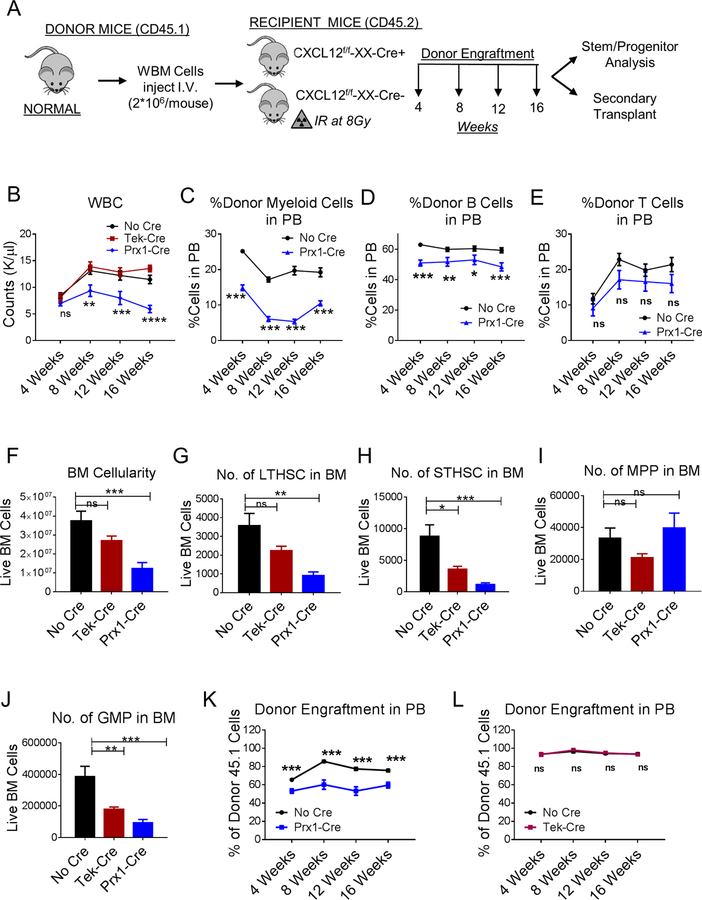
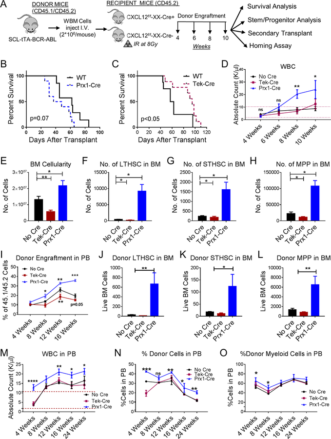
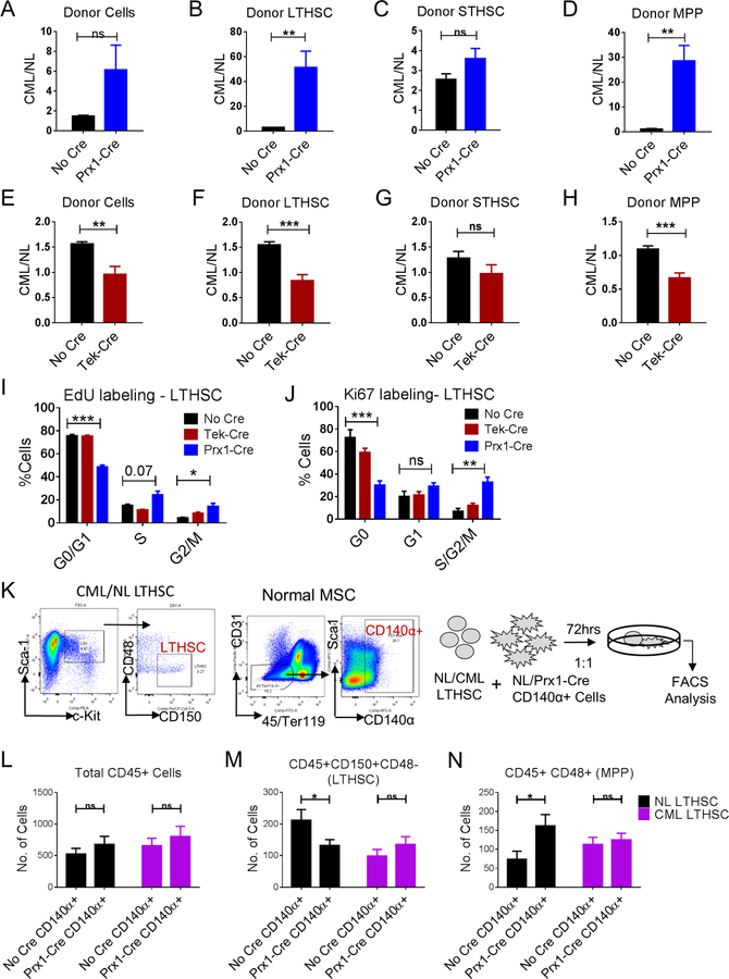
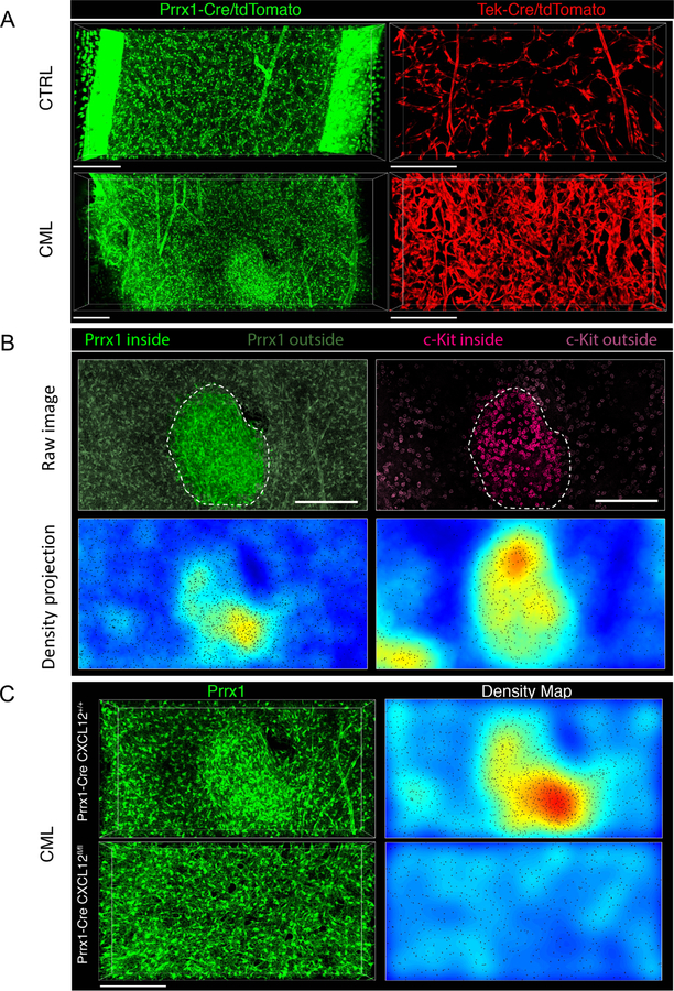
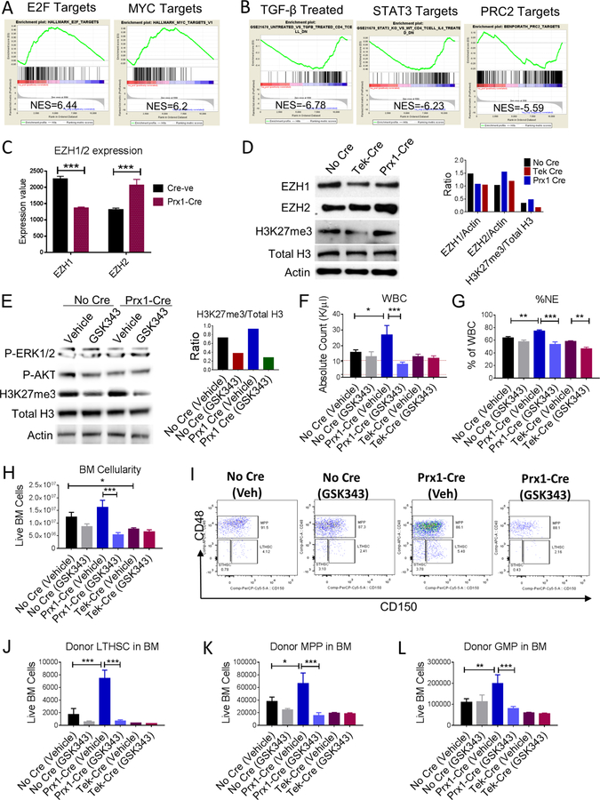
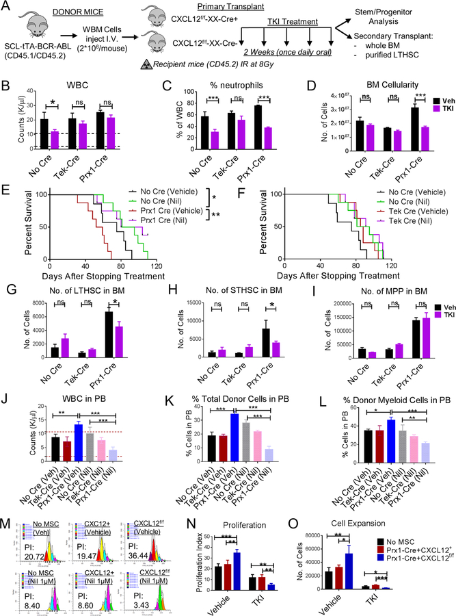
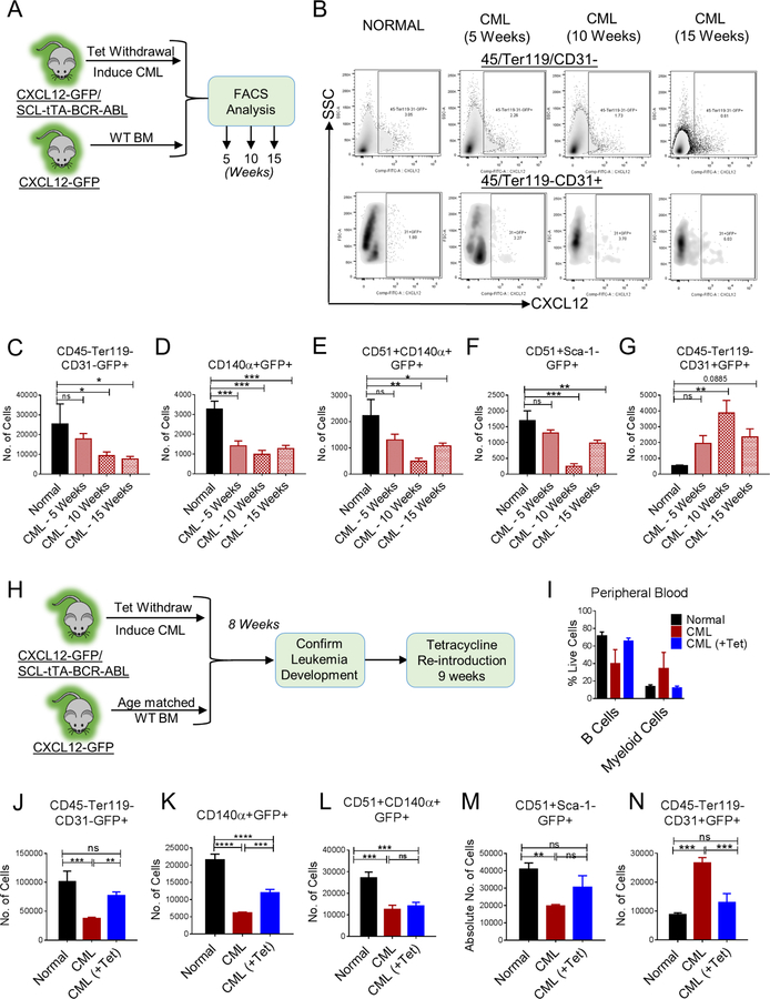
Chronic myeloid leukemia (CML) originates in a hematopoietic stem cell (HSC) transformed by the breakpoint cluster region (BCR)-abelson (ABL) oncogene and is effectively treated with tyrosine kinase inhibitors (TKIs). TKIs do not eliminate disease-propagating leukemic stem cells (LSCs), suggesting a deeper understanding of niche-dependent regulation of CML LSCs is required to eradicate disease. Cxcl12 is expressed in bone marrow niches and controls HSC maintenance, and here, we show that targeted deletion of Cxcl12 from mesenchymal stromal cells (MSCs) reduces normal HSC numbers but promotes LSC expansion by increasing self-renewing cell divisions, possibly through enhanced Ezh2 activity. In contrast, endothelial cell-specific Cxcl12 deletion decreases LSC proliferation, suggesting niche-specific effects. During CML development, abnormal clusters of colocalized MSCs and LSCs form but disappear upon Cxcl12 deletion. Moreover, MSC-specific deletion of Cxcl12 increases LSC elimination by TKI treatment. These findings highlight a critical role of niche-specific effects of Cxcl12 expression in maintaining quiescence of TKI-resistant LSC populations.

Keywords: CXCL12; TKI; bone marrow microenvironment; chronic myelogenous leukemia; drug resistance; hematopoietic stem cells; leukemia stem cells; mesenchymal stromal cells.


Conflict of interest statement

Declaration of Interests

The authors declare no competing financial interests.

Figures

Figure 1.
Figure 1.. CXCL12 produced from mesenchymal progenitor cells is important for normal murine LTHSC maintenance
(A) Whole bone marrow cells (WBM) were obtained from wild-type normal mice and injected (n=7–11 mice/group) into age and sex matched CXCL12f/f (CXCL12f/f-XX-Cre-; Cre-negative), CXCL12f/f-Tek-Cre, CXCL12f/f-Prx1-Cre, (CXCL12f/f-XX-Cre+; XX representing either Tek or Prx1) 6–8 week old littermates irradiated at 8Gy. Blood draws were performed every 4 weeks to check donor engraftment in PB until 16 weeks after which mice were euthanized to analyze stem/progenitor populations. PB white blood cells (WBC) (B), donor-derived myeloid cells (C), -B Cells (D), -T Cells (E), BM total cellularity (F), LTHSC (G), STHSC (H), MPP (I), GMP (J), per 1 femur, 1 tibia (2 bones) are shown. WBM cells from primary transplanted mice were pooled and transplanted into secondary recipient normal mice (n=3–10 mice/group) irradiated at 8Gy. Serial analysis for donor engraftment every 4 weeks is shown in the PB of mice receiving BM from either No Cre or Tek-Cre (K), and No Cre or Prx1-Cre mice (L). Error bars represent mean ± sem. Significance values. ns (non-significant) P>0.05, *P< 0.05, **P< 0.01, ***P<0.001, ****P<0.0001. See also Figures S1 and S2.
Figure 2.
Figure 2.. CXCL12 deletion from MSC and endothelial cells differentially regulates murine CML LSC maintenance and self-renewal
(A) BCR-ABL expression was induced in SCL-tTA-BCR-ABL mice by tetracycline withdrawal. Whole BM cells were obtained 6–8 weeks after induction and injected into age and sex matched CXCL12f/f (CXCL12f/f-XX-Cre-; Cre-negative), CXCL12f/f-Tek-Cre, CXCL12f/f-Prx1-Cre, (CXCL12f/f-XX-Cre+; XX representing either Tek or Prx1) 6–8-week old littermates irradiated at 800cGy (n=7–11 mice/group) (B-H). Survival of Prx1-Cre and Tek-Cre mice is shown (B, C). Blood draws were performed every 2 weeks to check for leukemia development. Total white blood cells (WBC) are shown (D). Mice were euthanized at 10 weeks to analyze stem/progenitor populations. BM cellularity (E), LTHSC (F), STHSC (G), and MPP (H) per 1 femur, 1 tibia (2 bones) are shown. In another experiment, CML c-Kit+ enriched cells (2*106 cells/mouse) were injected into non-irradiated anti-c-Kit and anti-CD47 antibodies treated recipient Cre mice. Serial CML donor engraftment in the PB (I), donor LTHSC (J), STHSC (K), MPP (L) in the BM are shown after 16 weeks. After primary transplantation of CML WBM cells into respective WT and knock out mice from experiment A, LTHSC were FACS purified and transplanted into secondary healthy WT mice irradiated at 8Gy. Serial blood draw was performed every 4 weeks until 24 weeks. The total WBC (M), frequency of donor engraftment (N) and frequency of donor myeloid cells in the PB (O) from secondary transplant experiment are shown. Error bars represent mean ± sem. Significance values. ns (non-significant) P>0.05, *P< 0.05, **P< 0.01, ***P<0.001, ****P<0.0001. See also Figures S3 and S4.
Figure 3.
Figure 3.. CXCL12 deletion from MSC results in increased CML LSC cycling and provides a competitive advantage to CML LTHSC
Ratio of donor total CML cells over normal cells (A), LTHSC (B), STHSC (C), MPP (D), in BM of either Cre-negative or Prx1-Cre recipient mice. Ratio of donor total CML cells over normal cells (E), LTHSC (F), STHSC (G), MPP (H), in BM of either Cre-negative or Tek-Cre recipient mice. Pooled cell cycle analysis of CML LTHSC from EdU incorporation and DAPI assay (I), and Ki67 and DAPI labeling (J). (K) LTHSC were FACS sorted from normal or CML mice, and CD140α+ cells were FACS sorted from normal or Prx1-Cre+ mice. Normal (CD45.2+) or CML (CD45.1+/CD45.2+) LTHSC were co-cultured with either normal (NL) or Prx1-Cre+ CD140α+ cells for 3 days and cell numbers and immunophenotype (CD45, CD150, CD48) were analyzed. The absolute number of total CD45+ cells (L), LTHSC (M), MPP (N), are shown. Error bars represent mean ± sem. Significance values. ns (non-significant) P>0.05, *P< 0.05, **P< 0.01, ***P<0.001, ****P<0.0001. See also Figure S5.
Figure 4.
Figure 4.. CXCL12 deletion prevents formation of MSC clusters in CML BM with colocalization of leukemic progenitors
Representative 3D confocal images of femoral BM volumes of Prx1-Cre+ Rosa26-tdTomatof/f (green) and Tek-Cre+ Rosa26-tdTomatof/f (red) mice transplanted with FACS sorted α-cat-GFP+ c-Kit+ cells from either normal (CTRL) or CML mice (CML) 8 weeks post-transplant. (A). Abnormal clusters of Prx1-Cre+ MSC appear in the BM during progression of CML (left panels: depth, 90 µm; size, 1429 µm*549 µm; scale bar, 200 µm; right panels: depth, 64 µm; size, 800 µm*400 µm; scale bar, 200 µm). CML progression has massive effects in BM microvascular structure compared to normal BM (right panels). A conspicuous increase in microvascular density, a loss of the typical sinusoidal morphology and prominent signs of vascular remodeling are apparent. (B) 3D images and tissue maps of typical MSC cluster and aggregation of c-Kit+ leukemic progenitors (magenta) inside and in the vicinity of these structures (delimited by dashed lines and highlighted) (top image: depth, 90.4 µm; size, 517 µm*526 µm; scale bar, 200 µm) (C) Representative images of MSC (green) in Prx1-Cre+ CXCL12+/+ (upper panel) and Prx1-Cre+ CXCL12fl/fl bones (lower panel) with CML, which shows normal homogeneous distribution of MSC and absence of clusters when CXCL12 is deleted from the mesenchymal Prx-1-Cre+ fraction (top left panel: depth, 107 µm; size, 800 µm*400 µm; scale bar, 200 µm; bottom left panel: depth, 64 µm; size, 800 µm*400 µm; scale bar, 200 µm). Right images show tissue density maps of MSC corresponding to BM volumes imaged in left panels. Automated microscopy images correspond to stitching of adjacent fields. See also Figure S6.
Figure 5.
Figure 5.. CXCL12 deletion from MSC results in downregulation of gene signatures associated with quiescence and TKI resistance in CML LSC
RNASeq analysis was performed on CML LTHSC FACS sorted from Cre-negative and Prx1-Cre mice. Gene-set Enrichment Analysis (GSEA) of differentially expressed genes showed that CML LTHSC from Prx1-Cre exhibited significantly increased expression of several gene sets related to cell cycling, hematopoietic stem cell proliferation, and MYC target genes when compared to LTHSC from Cre-negative mice. Representative plots for E2F target and MYC target genes are shown (A). CML LTHSC from Prx1-Cre showed decreased expression of gene sets related to TGF-β signaling, STAT3/5, and PRC2 target genes (B). (C) Expression of EZH1 and EZH2 among LTHSC from Prx1- Cre and Cre-negative mice from the RNA-Seq analysis. FACS sorted CML LSK cells from various groups of mice either untreated (D), or treated with vehicle or EZH2 inhibitor (GSK343) for 2 weeks and subjected to western blotting to analyze various protein levels (E) and densitometric analysis performed as shown. Effect of EZH2 inhibition on leukemia progression was assessed by analyzing total WBC levels (F), frequency of neutrophils in PB (G), BM cellularity (H), representative FACS plots showing CML LTHSC in BM (I), total no. of LTHSC (J), MPP (K), and GMP (L). Error bars represent mean ± sem. Significance values. ns (non-significant) P>0.05, *P< 0.05, **P< 0.01, ***P<0.001, ****P<0.0001. See also Table S1.
Figure 6.
Figure 6.. CXCL12 deletion from MSC sensitizes murine and human CML LSC to TKI treatment
(A) BCR-ABL expression was induced in SCL-tTA-BCR-ABL mice by tetracycline withdrawal. Whole BM cells were obtained 6–8 weeks after induction and injected into age and sex matched CXCL12f/f (CXCL12f/f-XX-Cre-; Cre-negative), CXCL12f/f-Tek-Cre, CXCL12f/f-Prx1-Cre, (CXCL12f/f-XX-Cre+; XX representing either Tek or Prx1) 6–8-week old littermates irradiated at 8Gy (n=5–6 mice/group). 8 weeks post-transplantation, mice were treated with either vehicle (Veh) or nilotinib (50mg/kg; TKI) for 2 weeks once daily oral gavage after which mice were euthanized and PB, BM, spleen analyzed. Total white blood cells (WBC) (B) and frequency of neutrophils (C) in the PB are displayed. A cohort of mice were followed for their survival after stopping treatment among No Cre and Prx1-Cre mice (E), and No Cre and Tek-Cre mice (F). BM LTHSC (G), STHSC (H), MPP (I) per 1 femur, 1 tibia (2 bones) are shown. FACS sorted LTHSC from primary vehicle or TKI-treated mice (CD45.1/2+) were pooled and transplanted into secondary recipient (CD45.2+) healthy WT 6–8-week-old mice (1000 cells/mouse+250,000 helper WBM CD45.2+ cells) irradiated at 8Gy. Long-term donor engraftment was performed 16 weeks following transplantation. The total WBC levels (J), long-term CML donor engraftment (K), frequency of donor myeloid cells (L), in the PB of mice are shown. Human CML 34+ cells were stained with CFSE. CFSE+ primitive cells (34+ 38-) were FACS sorted and cultured in the presence or absence of FACS sorted tdTomato+ cells from CXCL12+/+-tdTomatof/+-Prx1-Cre+ and CXCL12f/f-tdTomatof/+-Prx1-Cre+ mice for 3 days and treated with or without Nil (1µM). Representative FACS plot (M), proliferation (N) and expansion (O) are shown. Error bars represent mean ± sem. ns (non-significant) P>0.05, *P< 0.05, **P< 0.01, ***P<0.001, ****P<0.0001. See also Figure S7.
Figure 7.
Figure 7.. CML development leads to altered distribution of CXCL12-expressing cells in murine BM
(A) Age and sex matched CXCL12-GFP knock-in mice (Normal) and CXCL12-GFP-SCL-tTA-BCR-ABL mice (CML) were subjected to tetracycline withdrawal. Mice were euthanized and FACS analysis for BM microenvironmental cells was performed every 5 weeks until 15 weeks (n=4–5 mice/group). FACS plots for CXCL12-expressing stromal (45-Ter119-CD31-) and endothelial cells (45-Ter119-CD31+) during CML development are shown (B). The number of CXCL12-expressing stromal cells (CD45-Ter119-CD31-GFP+) (C), CXCL12-expressing PDGFRα+ cells (CD45-Ter119-CD31-CD140α+GFP+) (D), CXCL12-expressing osteoprogenitors (CD45-Ter119-CD31-CD51+CD140α+GFP+) (E), CXCL12-expressing osteoblasts (CD45-Ter119-CD31-CD51+Sca-1-GFP+) (F), and CXCL12-expressing endothelial cells (CD45/Ter119-CD31+GFP+) (G) per femur are shown. Error bars represent mean ± sem. Significance values. ns (non-significant) P>0.05, *P< 0.05, **P< 0.01, ***P<0.001, ****P<0.0001. (H) Age and sex matched CXCL12-GFP knock-in mice (Normal) and CXCL12-GFP-SCL-tTA-BCR-ABL mice (CML) were subjected to tetracycline withdrawal for 8 weeks to induce leukemia. Then, a cohort of CML mice were placed back on tetracycline for further 9 weeks (CML+Tet). (I) Multilineage blood profile showing restoration of normal hematopoiesis upon tetracycline re-introduction. Mice were euthanized and FACS analysis was performed on BM (n=3–4 mice/group). The number of CXCL12-expressing stromal cells (CD4-Ter119-CD31-GFP+) (J), CXCL12-expressing PDGFRα+ cells (CD45-Ter119-CD31-CD140α+GFP+) (K), CXCL12-expressing osteoprogenitors (CD45-Ter119-CD31-CD51+CD140α+GFP+) (L), CXCL12-expressing osteoblasts (CD45-Ter119-CD31-CD51+Sca-1-GFP+) (M), and CXCL12-expressing endothelial cells (CD45-Ter119-CD31+GFP+) (N) per femur are shown. Error bars represent mean ± sem. Significance values. ns (non-significant) P>0.05, *P< 0.05, **P< 0.01, ***P<0.001, ****P<0.0001.

Comment in

References

    1. Abe-Suzuki S, Kurata M, Abe S, Onishi I, Kirimura S, Nashimoto M, Murayama T, Hidaka M, and Kitagawa M (2014). CXCL12+ stromal cells as bone marrow niche for CD34+ hematopoietic cells and their association with disease progression in myelodysplastic syndromes. Laboratory investigation; a journal of technical methods and pathology 94, 1212–1223. - PubMed
    1. Abraham M, Klein S, Bulvik B, Wald H, Weiss ID, Olam D, Weiss L, Beider K, Eizenberg O, Wald O, et al. (2017). The CXCR4 inhibitor BL-8040 induces the apoptosis of AML blasts by downregulating ERK, BCL-2, MCL-1 and cyclin-D1 via altered miR-15a/16–1 expression. Leukemia - PubMed
    1. Acar M, Kocherlakota KS, Murphy MM, Peyer JG, Oguro H, Inra CN, Jaiyeola C, Zhao Z, Luby-Phelps K, and Morrison SJ (2015). Deep imaging of bone marrow shows non-dividing stem cells are mainly perisinusoidal. Nature 526, 126–130. - PMC - PubMed
    1. Agarwal P, Zhang B, Ho Y, Cook A, Li L, Mikhail FM, Wang Y, McLaughlin ME, and Bhatia R (2016). Enhanced targeting of CML stem and progenitor cells by inhibition of porcupine acyltransferase in combination with TKI. Blood - PMC - PubMed
    1. Anders S, Pyl PT, and Huber W (2015). HTSeq--a Python framework to work with high-throughput sequencing data. Bioinformatics 31, 166–169. - PMC - PubMed

Publication types

MeSH terms