. 2019 Apr:42:296-303.
doi: 10.1016/j.ebiom.2019.03.020. Epub 2019 Mar 21.

Genomic landscape of allelic imbalance in premalignant atypical adenomatous hyperplasias of the lung

Affiliations

Genomic landscape of allelic imbalance in premalignant atypical adenomatous hyperplasias of the lung

Smruthy Sivakumar et al. EBioMedicine. 2019 Apr.

Abstract

Background: Genomic investigation of atypical adenomatous hyperplasia (AAH), the only known precursor lesion to lung adenocarcinomas (LUAD), presents challenges due to the low mutant cell fractions. This necessitates sensitive methods for detection of chromosomal aberrations to better study the role of critical alterations in early lung cancer pathogenesis and the progression from AAH to LUAD.

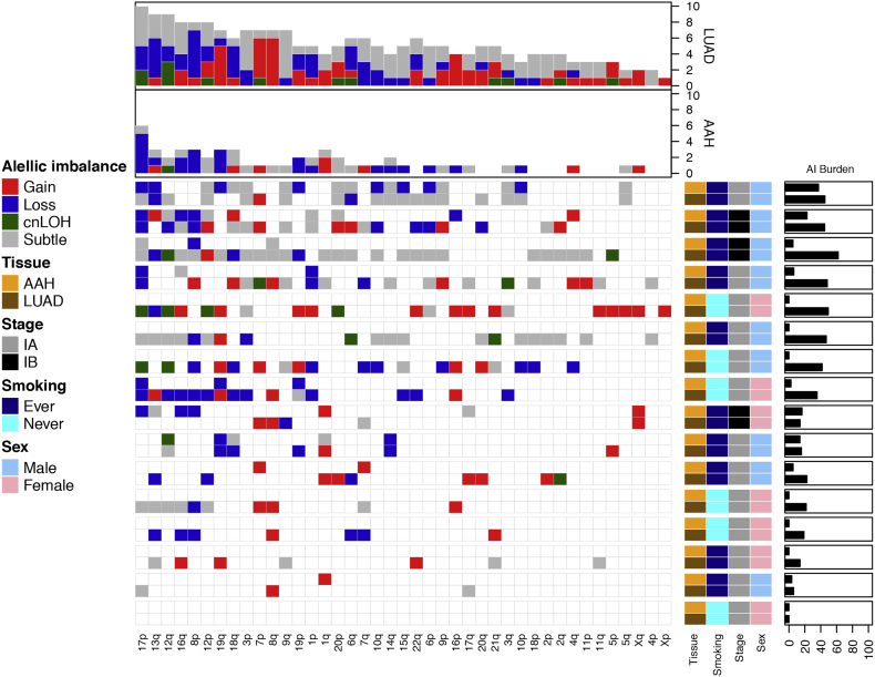
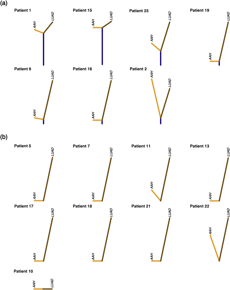
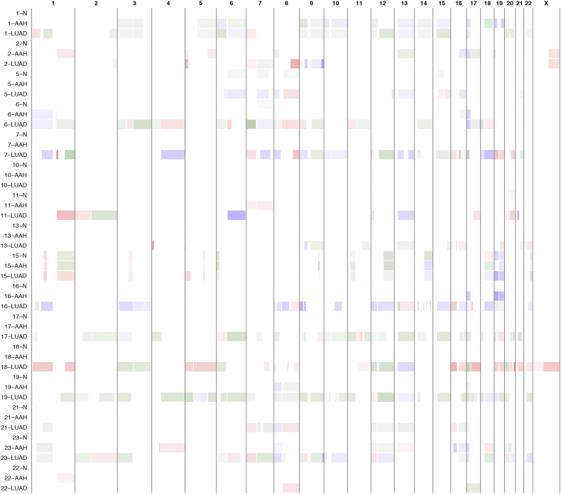
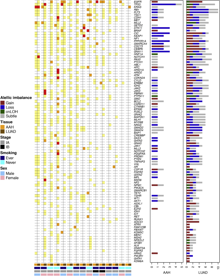
Methods: We applied a sensitive haplotype-based statistical technique to detect chromosomal alterations leading to allelic imbalance (AI) from genotype array profiling of 48 matched normal lung parenchyma, AAH and tumor tissues from 16 stage-I LUAD patients. To gain insights into shared developmental trajectories among tissues, we performed phylogenetic analyses and integrated our results with point mutation data, highlighting significantly-mutated driver genes in LUAD pathogenesis.

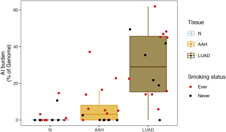
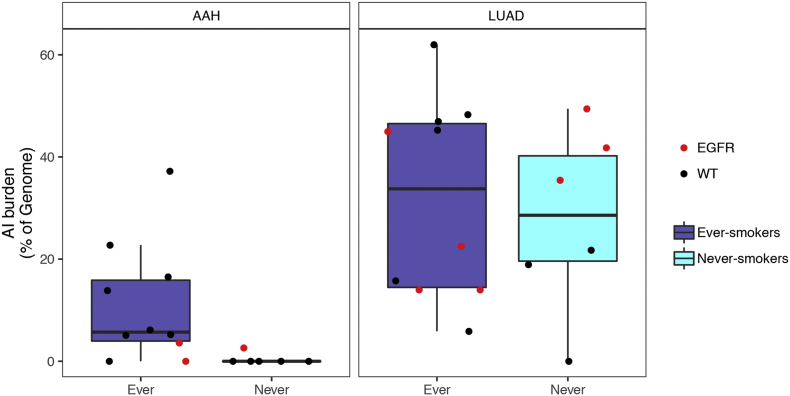
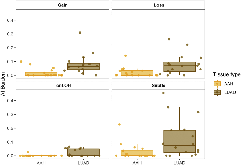
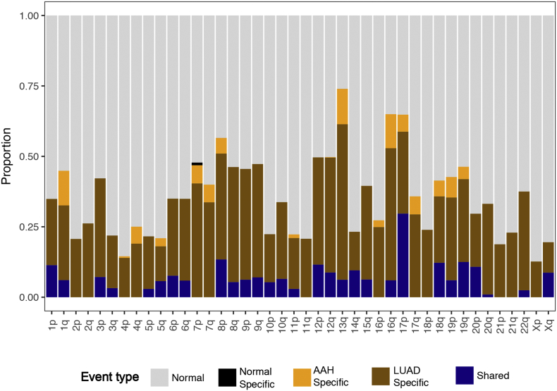
Findings: AI was detected in nine AAHs (56%). Six cases exhibited recurrent loss of 17p. AI and the enrichment of 17p events were predominantly identified in patients with smoking history. Among the nine AAH tissues with detected AI, seven exhibited evidence for shared chromosomal aberrations with matched LUAD specimens, including losses harboring tumor suppressors on 17p, 8p, 9p, 9q, 19p, and gains encompassing oncogenes on 8q, 12p and 1q.

Interpretation: Chromosomal aberrations, particularly 17p loss, appear to play critical roles early in AAH pathogenesis. Genomic instability in AAH, as well as truncal chromosomal aberrations shared with LUAD, provide evidence for mutation accumulation and are suggestive of a cancerized field contributing to the clonal selection and expansion of these premalignant lesions. FUND: Supported in part by Cancer Prevention and Research Institute of Texas (CPRIT) grant RP150079 (PS and HK), NIH grant R01HG005859 (PS) and The University of Texas MD Anderson Cancer Center Core Support Grant.

Keywords: Allelic imbalance; Atypical adenomatous hyperplasia; Chromosomal instability; Lung adenocarcinoma; Pathogenesis; Preneoplasia.


Figures

Fig. 1
Fig. 1
Chromosomal allelic imbalance burden in normal, AAH and LUAD tissues. Regions with subtle chromosomal allelic imbalance (AI) were identified in the normal (N), AAH and matched LUAD tissues using genome-wide genotype array profiling as described in the Methods section. AI burdens, defined as a percent of the genome, are represented by box plots for each tissue type (N, AAH and LUAD). The burden for each patient is shown as a point overlaid on the boxplots. The points are colored red if the patient had a smoking history and black if the patient was a non-smoker. (For interpretation of the references to colour in this figure legend, the reader is referred to the web version of this article.)
Fig. 2
Fig. 2
Genome-wide chromosomal arm allelic imbalance events in matched AAH and LUAD. We identified 53 subtle chromosomal arm events in AAHs and 210 chromosomal arm events in matched LUADs across 16 stage-I LUAD patients. The distribution and type of these events are shown, with rows representing individual patients and columns representing chromosome arms. Each individual row is further divided to show profiles of matched AAH and LUAD from that individual. The events are annotated as gain (red), loss (blue) or copy-neutral loss of heterozygosity (cnLOH, green) while unclassifiable events are annotated as subtle (gray). The overall burden across all chromosomal arms is shown in the bar plots at the top, while allelic imbalance burdens in each sample are shown on the right. Patients are also annotated to denote their clinicopathological features. (For interpretation of the references to colour in this figure legend, the reader is referred to the web version of this article.)
Fig. 3
Fig. 3
Phylogenetic reconstruction of truncal, AAH-specific and LUAD-specific chromosomal aberrations. Matched AAH and LUAD specimens from individual patients were assessed for patterns of shared as well as tissue-specific allelic imbalance events and phylogenetic rooted trees were constructed as described in the Methods section. Cases exhibiting any evidence for shared events are shown in (a) and remaining cases are shown in (b). Vertical distances in each tree are scaled to the proportion of shared as well as tissue-specific events. Shared events, thereby trunks of the trees, are shown in dark blue; while tissue-specific events are shown separately for AAH (orange) and LUAD (brown). (For interpretation of the references to colour in this figure legend, the reader is referred to the web version of this article.)
Supplementary Fig. 1
Chromosomal allelic imbalance burden in AAH and LUAD based on tobacco history. Identification of allelic imbalance (AI) and estimation of genomic AI burden was performed as described in the Methods section. AI burdens, defined as a percent of the genome, are represented by box plots for each tissue type (AAH and LUAD) based on tobacco history (never-smoker and ever-smoker). The burdens for each individual case are overlaid as points on the boxplot. Specifically, samples exhibiting EGFR mutations are shown as red dots.
Supplementary Fig. 2
Chromosomal allelic imbalance burden in AAH and LUAD for each event type. Regions with chromosomal allelic imbalance (AI) were identified in AAH and matched LUADs and classified into gain, loss and copy-neutral loss of heterozygosity (cnLOH), from genome-wide genotype array profiling as described in the Methods section. The remaining unclassifiable events are shown as “subtle”. AI burdens, defined as a percent of the genome aberrant, are represented by tissue-level box plots (AAH and LUAD) for each event category. The burdens for each individual case are overlaid as points on the boxplot.
Supplementary Fig. 3
Chromosomal arm and focal allelic imbalance events in matched normal lung parenchyma, AAH and LUAD. The genomic locations of the identified chromosomal allelic imbalance events were plotted for all 48 samples of matched normal lung parenchyma, AAH and LUAD from 16 patients. Allelic imbalance regions are first classified as gains (red) or losses (blue) as described in the Methods section, the intensity of which is based on the log R ratio of the event. The remaining events are annotated in green as subtle and copy-neutral loss of heterozygosity (cnLOH) events, intensity of which is based on B-allele frequency deviation for the event region, with darker shaded regions representing increased evidence for cnLOH.
Supplementary Fig. 4
Distribution of shared and tissue-specific allelic imbalance events across chromosomal arms. The identified allelic imbalance (AI) events across all patients were averaged and assessed for chromosomal patterns of shared as well as tissue-specific events. A stacked bar plot representing the proportion of normal region (gray), shared AI (blue), AAH-specific AI (orange), LUAD-specific AI (brown) and normal tissue-specific AI (black) for each chromosome arm is shown.
Supplementary Fig. 5
Progressive and somatic two-hit processes in matched AAH and LUADs. Known cancer driver genes [[11], [12]] within regions of allelic imbalance or with single nucleotide mutations (SNVs) [6] were examined. The figure depicts genes exhibiting somatic “two-hits” (both SNVs and AI; red) in AAHs and LUADs as well as those exhibiting a first shared hit (either SNV: orange or AI: yellow) in the AAH accompanied by a second tumor-specific hit in the matched LUAD. For samples with allelic imbalance, the event types are shown as bar plots on the right, accompanying each gene, in both the AAH and LUAD specimens.

Comment in

References

    1. Herbst R.S., Heymach J.V., Lippman S.M. Lung cancer. N Engl J Med. 2008;359:1367–1380. - PMC - PubMed
    1. Kadara H., Scheet P., Wistuba I.I., Spira A.E. Early events in the molecular pathogenesis of lung cancer. Cancer Prev Res. 2016;9:518–527. - PubMed
    1. Negrini S., Gorgoulis V.G., Halazonetis T.D. Genomic instability — an evolving hallmark of cancer. Nat Rev Mol Cell Biol. 2010;11:220–228. - PubMed
    1. Takamochi K., Ogura T., Suzuki K., Kawasaki H., Kurashima Y., Yokose T. Loss of heterozygosity on chromosomes 9q and 16p in atypical adenomatous hyperplasia concomitant with adenocarcinoma of the lung. Am J Pathol. 2001;159:1941–1948. - PMC - PubMed
    1. Wistuba I.I., Gazdar A.F. Lung cancer preneoplasia. Annu Rev Pathol. 2006;1:331–348. - PubMed

MeSH terms