. 2019 Jan-Feb;53(1):128-137.
doi: 10.4103/ortho.IJOrtho_631_17.

When and How to Operate Fragility Fractures of the Pelvis?

Affiliations

When and How to Operate Fragility Fractures of the Pelvis?

Pol Maria Rommens et al. Indian J Orthop. 2019 Jan-Feb.

Abstract

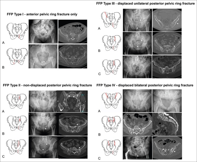
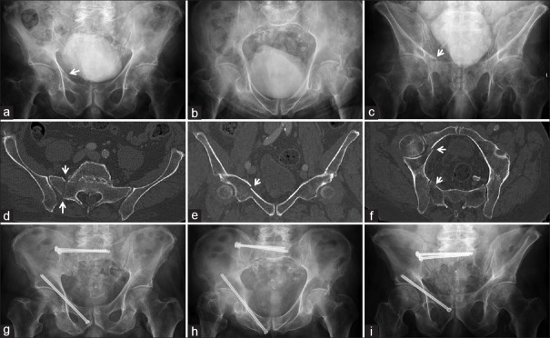
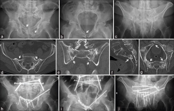
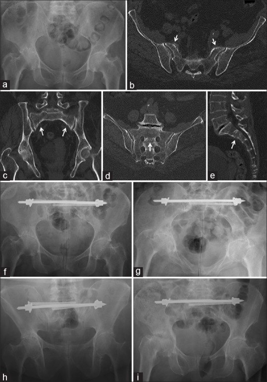
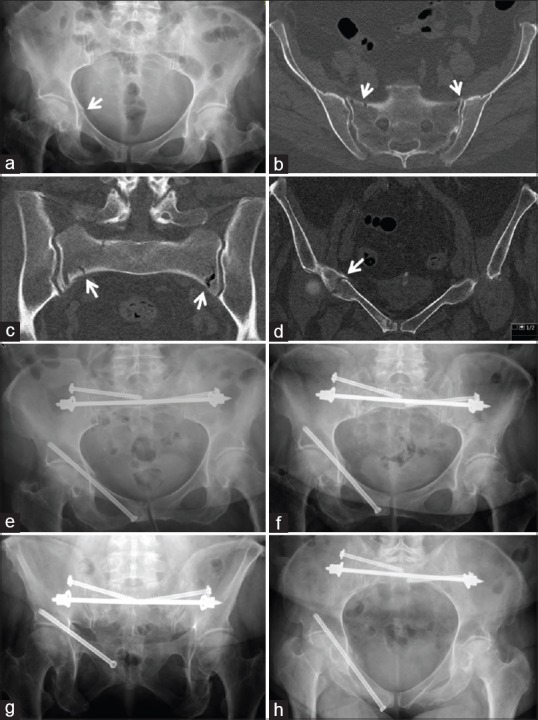
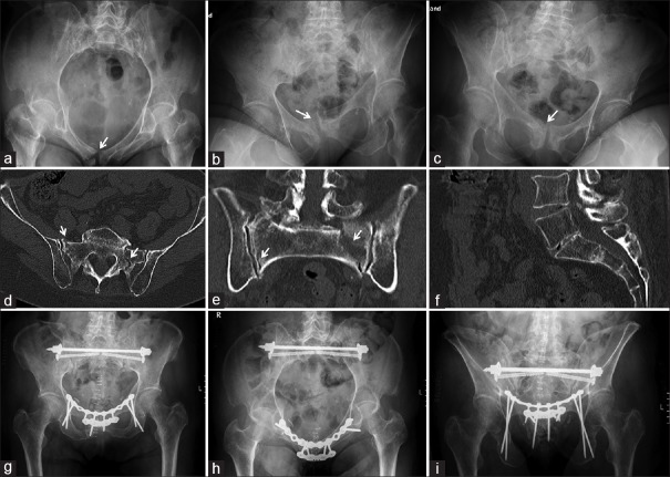
Fragility fractures of the pelvis (FFP) are an entity with an increasing frequency. The characteristics of these fractures are different from pelvic ring fractures in younger adults. There is a low energy instead of a high energy trauma mechanism. Due to a specific and consistent decrease of bone mineral density, typical fractures in the anterior and posterior pelvic ring occur. Bilateral sacral ala fractures are frequent. A new classification system distinguishes between four categories with increasing loss of stability. The subtypes represent different localizations of fractures. The primary goal of treatment is restoring mobility and independency. Depending on the amount of instability, conservative or surgical treatment is recommended. The operative technique should be as less invasive as possible. When the broken posterior pelvic ring is fixed operatively, a surgical fixation of the anterior pelvic ring should be considered as well. FFP Type I can be treated conservatively. In many cases, FFP Type II can also be treated conservatively. When conservative treatment fails, percutaneous fixation is performed. FFP Type III and FFP Type IV are treated operatively. The choice of the operation technique is depending on the localization of the fracture. Iliosacral screw osteosynthesis, transsacral bar osteosynthesis, transiliac internal fixation, and iliolumbar fixation are alternatives for stabilization of the posterior pelvic ring. Plate osteosynthesis, retrograde transpubic screw, and anterior internal fixation are alternatives for stabilization of the anterior pelvic ring. Postoperatively, early mobilization, with weight bearing as tolerated, is started. Simultaneously, bone metabolism is also analyzed and its defects compensated. Medical comorbidities should be identified and treated with the help of a multidisciplinary team.

Keywords: Characteristics; classification; fragility fractures pelvis; geriatric comanagement; minimally invasive; surgical fixation.


Conflict of interest statement

There are no conflicts of interest.

Figures

Figure 1
Figure 1
Classification of fragility fractures of the pelvis
Figure 2
Figure 2
A 70-year-old male suffered a fall. (a) AP pelvic X-ray: right pubic ramus fracture (arrow). (b) Pelvic inlet. (c) Pelvic outlet: pubic ramus fracture (arrow). (d) Axial computed tomography: fracture of the right sacral ala (arrows). (e) Coronal computed tomography: superior pubic ramus fracture (arrow). (f) Oblique computed tomography reconstruction: fractures at anterior and posterior pelvis (arrows). It concerns a fragility fractures of the pelvis Type IIc. (g) AP pelvic X-ray after 3 months. Sacral fracture was stabilized with two iliosacral screws, pubic ramus fracture with long retrograde transpubic screw. (h) Pelvic inlet. (i) Pelvic outlet
Figure 3
Figure 3
A 76-year-old male with pelvic irradiation after prostatectomy. (a) AP pelvic X-ray: bilateral pubic ramus fractures (arrows). (b) Pelvic inlet. (c) Pelvic outlet. (d) Axial computed tomography: bilateral sacral ala fractures (arrows). (e) Coronal computed tomography. (f) Sagittal computed tomography: horizontal fracture between S2 and S3. It concerns a fragility fractures of the pelvis Type IVb. (g) Oblique computed tomography with all fractures. (h) Postoperative pelvic X-ray. Transiliac internal fixation and two iliosacral screws for the posterior pelvis, retrograde transpubic screw for the left, plate and screws for the right anterior fracture. (i) Pelvic inlet. (j) Pelvic outlet
Figure 4
Figure 4
A 65-year-old female with 8 months of intense pain. (a) Normal AP pelvis X-ray. (b) Axial computed tomography: bilateral sacral ala fractures (arrows). (c) Coronal computed tomography: bilateral sacral ala fractures (arrows). (d) Oblique computed tomography: Fracture between left and right S1-neuroforamen (arrow). (e) Sagittal midline computed tomography: horizontal fracture between S1 and S2 (arrow). It concerns a fragility fractures of the pelvis Type IVb. (f) Postoperative AP pelvic X-ray. Fractures were stabilized with transsacral bar and two iliosacral screws. (g) Pelvic inlet. (h) Pelvic outlet. (i) AP pelvic X-ray 2 years later showing the right acetabular fracture
Figure 5
Figure 5
A 75-year-old female suffered a domestic fall. (a) AP pelvic X-ray: right superior pubic ramus fracture. (b) Transverse computed tomography: bilateral sacral ala fractures. (c) Coronal computed tomography: bilateral sacral ala fracture. (d) Coronal computed tomography: superior pubic ramus fracture on the right. It concerns a fragility fracture of the pelvis Type IIC. (e) Postoperative AP pelvic X-ray: transsacral bar, two iliosacral screws, and retrograde transpubic screw. (f) Pelvic inlet. (g) Pelvic outlet. (h) AP pelvic X-ray after 3 months. Bridging callus at the pubic ramus fracture
Figure 6
Figure 6
A 74-year-old female with pelvic pain. (a) AP pelvic X-ray: right pubic bone fracture (arrow). (b) Right one-leg stand: vertical instability of pubic symphysis (arrow). (c) Left one-leg stand confirms instability (arrow). (d) Axial computed tomography: bilateral sacral ala fractures (arrows). (e) Coronal computed tomography: same fractures. (f) Mid-sagittal computed tomography: normal. (g) Postoperative AP pelvic X-ray: transsacral bar and two iliosacral screws posteriorly, two plates and screws anteriorly. Marginal screws of the upper plate use infraacetabular corridor, anterior plate is angular stable. (h) Pelvic inlet. (i) Pelvic outlet

References

    1. Hernlund E, Svedbom A, Ivergård M, Compston J, Cooper C, Stenmark J, et al. Osteoporosis in the European union: Medical management, epidemiology and economic burden. A report prepared in collaboration with the International Osteoporosis Foundation (IOF) and the European Federation of Pharmaceutical Industry Associations (EFPIA) Arch Osteoporos. 2013;8:136. - PMC - PubMed
    1. Johnell O, Kanis JA. An estimate of the worldwide prevalence and disability associated with osteoporotic fractures. Osteoporos Int. 2006;17:1726–33. - PubMed
    1. Watts NB. The fracture risk assessment tool (FRAX®): Applications in clinical practice. J Womens Health (Larchmt) 2011;20:525–31. - PubMed
    1. Rommens PM, Wagner D, Hofmann A. Surgical management of osteoporotic pelvic fractures: A new challenge. Eur J Trauma Emerg Surg. 2012;38:499–509. - PMC - PubMed
    1. Sullivan MP, Baldwin KD, Donegan DJ, Mehta S, Ahn J. Geriatric fractures about the hip: Divergent patterns in the proximal femur, acetabulum, and pelvis. Orthopedics. 2014;37:151–7. - PubMed

LinkOut - more resources