. 2019 Apr;17(4):2407-2416.
doi: 10.3892/etm.2019.7251. Epub 2019 Feb 7.

Assessing biological aging following systemic administration of bFGF-supplemented adipose-derived stem cells with high efficacy in an experimental rat model

Affiliations

Assessing biological aging following systemic administration of bFGF-supplemented adipose-derived stem cells with high efficacy in an experimental rat model

Hahn-Sol Bae et al. Exp Ther Med. 2019 Apr.

Abstract

Biological aging (BA) is a tool for comprehensive assessment of individual health status. A rat model was developed for measuring BA by intravenously administering adipose-derived stem cells (ADSCs) into rats and evaluating several biochemical parameters. In addition, the effect of basic fibroblast growth factor (bFGF) on the differentiation potential of ADSCs was analyzed. A total of 12 male Sprague Dawley rats were divided into autologous ADSC administration (n=6) and saline administration (n=6) groups. The ADSC administration group was further divided into the bFGF supplemented (n=3) and bFGF non-supplemented (n=3) groups. Biochemical parameters and antioxidant potential were evaluated prior to fat harvest and ADSC administration, as well as 1, 3, and 5 weeks following ADSC administration. ADSC administration regulated inflammation, renal and hepatic functions, and levels of antioxidant enzymes. The cell doubling time of the bFGF-supplemented group was shorter (P=0.0001) than that of the bFGF non-supplemented group. Renal and hepatic functions were maintained with bFGF supplementation, which possibly enhanced the effect of ADSCs. The rat model developed in the present study may promote better understanding of BA in the context of bFGF-supplemented ADSC administration.

Keywords: adipose-derived stem cell; basic fibroblast growth factor; biochemical parameter; biologic aging; rat model.


Figures

Figure 1.
Figure 1.
The schematic schedule of the present experimental protocol. The isolated fat tissues were washed and digested. A total of 106 ADSCs (P2) were administered via the femoral vein. At fat harvest, ADSC injection, and 1,3, and 5 weeks later, blood and urine samples were analyzed. ADSC, adipose-derived stem cells; bFGF, basic fibroblast growth factor.
Figure 2.
Figure 2.
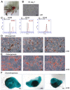
(A) Harvesting autologous inguinal fat pads from each rat. Following shaving and draping of the left inguinal region, a 2.0-cm incision was made along the inguinal fold, and the autologous inguinal fat pads were harvested. (B) Characterization of ADSCs. An optical microscope view of cultured ADSCs was acquired (magnification, ×40). (C) Flow cytometry analysis with common stem cell markers CD90 and CD44 and hematopoietic marker CD45 for characterizing autologous ADSCs prior to administration. (D) Alizarin red S stained cells underwent osteogenic differentiation. (E) Oil red O-positive lipid droplets indicate that cells underwent adipogenic differentiation. (F) Alcian blue staining was performed to detect chondrogenic differentiation. ADSC; adipose-derived stem cells; CD, cluster of differentiation; FITC, fluorescein isothiocyanate.
Figure 3.
Figure 3.
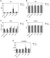
Alterations in leukocyte variables and serum or urine kidney signal. Blood and urine were obtained from the rat model each time-point, and complete blood count and chemical analyses were performed. Measurements in lean control and with ADSC administration were evaluated for (A) blood monocytes, (B) MPV, (C) serum BUN, (D) serum creatinine, and (E) UCCR are presented. The value of monocytes was sustained in ADSC injected group compared with that in saline group, which was increased. Conversely, all others (MPV, BUN, creatinine and UCCR) were downregulated following ADSC administration. Data are presented as the mean ± standard error of the mean. ADSC; adipose-derived stem cells; MPV, mean platelet volume; BUN, blood urea nitrogen; UCCR, urine cortisol:creatinine ratio.
Figure 4.
Figure 4.
Serum cholesterol, SOD, calcium, and α-amylase levels were determined. Bar graphs presenting (A) LDL, (B) HDL, (C) SOD, (D) calcium and (E) α-amylase levels. Serum LDL and serum HDL levels were downregulated in the ADSC administration group, which tends to reduce the risk of heart diseases. SOD level was increased following ADSC administration. Serum calcium level was lower in the ADSC administration group in a time-dependent manner. The level of α-amylase, a calcium metalloenzyme, was also lower following ADSC administration, thus ADSC can maintain homeostasis of the blood glucose level. Data are presented as the mean ± standard error of the mean. SOD, superoxide dismutase; LDL, low-density lipoprotein; HDL, high-density lipoprotein; ADSC; adipose-derived stem cells.
Figure 5.
Figure 5.
Doubling time of ADSCs cultured with or without bFGF. bFGF markedly increased the cell doubling rate at P1 and P2. Doubling time was markedly shorter in the bFGF treated group, thus indicating the progenitor cell feature. Data are presented as the mean ± standard error of the mean. *P<0.0001. ADSC; adipose-derived stem cells; bFGF, basic fibroblast growth factor; P1, passage 1; P2, passage 2.
Figure 6.
Figure 6.
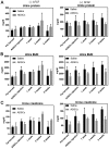
The liver signal can be regulated by administration of ADSCs cultured with bFGF supplementation. (A) ALT and (B) α-amylase levels in bFGF supplemented (right) and non-supplemented (left) groups. ALT level was downregulated in the bFGF-supplemented ADSC administration group. Additionally, α-amylase level was unchanged in bFGF non-treated group, but it was lower in the bFGF-supplemented group. This suggested that ADSCs maintain liver metabolism in anti-aging systems. Data are presented as the mean ± standard error of the mean. ADSC; adipose-derived stem cells; bFGF, basic fibroblast growth factor; ALT, alanine transaminase.
Figure 7.
Figure 7.
The markers of kidney function changed following ADSC administration in the bFGF supplemented group. (A) Urine protein, (B) urine BUN, and (C) urine creatinine levels in bFGF supplemented (right) and non-supplemented (left) groups. Urine protein was markedly decreased in the bFGF-supplemented ADSC administration group compared with bFGF non-treated group. Urine BUN and creatinine levels exhibited similar patterns to that of urine protein. bFGF affects the biological markers associated with hepatic and renal functions. Data are presented as the mean ± standard error of the mean. ADSC; adipose-derived stem cells; bFGF, basic fibroblast growth factor; BUN, blood urea nitrogen.

Similar articles

Cited by

References

    1. World Population Prospects: The 2017 Revision. Executive Summary. United Nations Department of Economic and Social Affairs, Population Division. https://esa.un.org/unpd/wpp/publications/files/wpp2017_keyfindings.pdf. [Jun 21;2017 ];2017
    1. Klemera P, Doubal B. A new approach to the concept and computation of biological age. Mech Ageing Dev. 2006;127:204–248. doi: 10.1016/j.mad.2005.10.004. - DOI - PubMed
    1. Piantanelli L, Rossolini G, Basso A, Piantanelli A, Malavolta M, Zaia A. Use of mathematical models of survivorship in the study of biomarkers of aging: The role of heterogeneity. Mech Ageing Dev. 2001;122:1461–1475. doi: 10.1016/S0047-6374(01)00271-8. - DOI - PubMed
    1. Jackson S, Weale MR, Weale RA. Biological age-what is it and can it be measured? Arch Gerontol Geriatr. 2003;36:103–115. doi: 10.1016/S0167-4943(02)00060-2. - DOI - PubMed
    1. Nakamura E, Lane MA, Roth GS, Ingram DK. A strategy for identifying biomarkers of aging: Further evaluation of hematology and blood chemistry data from a calorie restriction study in rhesus monkeys. Exp Gerontol. 1998;33:421–443. doi: 10.1016/S0531-5565(97)00134-4. - DOI - PubMed