. 2019 Apr;17(4):2855-2860.
doi: 10.3892/etm.2019.7230. Epub 2019 Jan 31.

CD147 mediates transforming growth factor-β1-induced epithelial-mesenchymal transition and cell invasion in squamous cell carcinoma of the tongue

Affiliations

CD147 mediates transforming growth factor-β1-induced epithelial-mesenchymal transition and cell invasion in squamous cell carcinoma of the tongue

Shinsuke Suzuki et al. Exp Ther Med. 2019 Apr.

Abstract

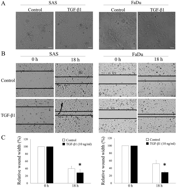
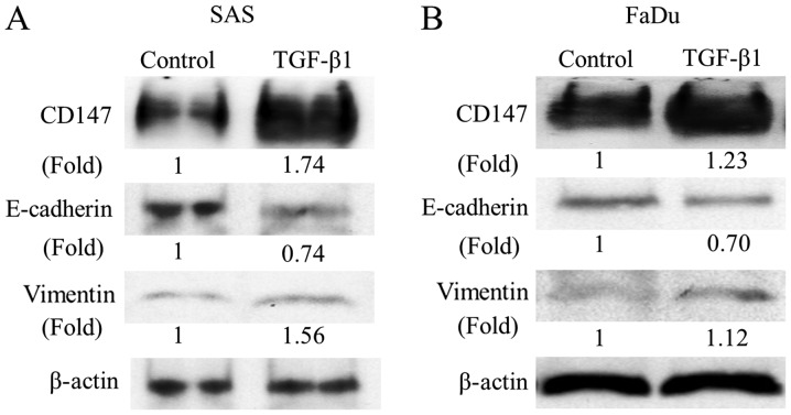
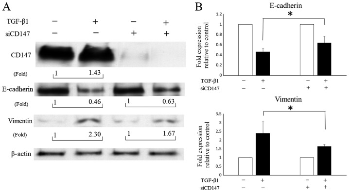
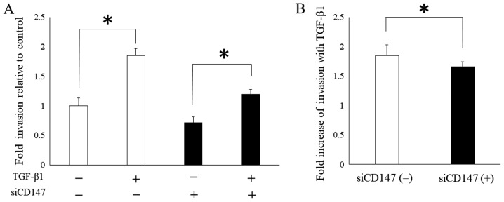
Epithelial-mesenchymal transition (EMT) is a physiological process in which epithelial cells attain the motile and invasive characteristics of mesenchymal cells, which results in the development of increased migratory and invasive cell behavior, serving as a vital mechanism of cancer progression. Hence, controlling the EMT for cancer treatment, including head and neck squamous cell carcinoma (HNSCC), is imperative. Among EMT-associated factors, transforming growth factor-β (TGF-β) is a well-established potent inducer. Recent research has revealed that CD147, a member of the immunoglobulin superfamily, promotes the EMT. However, the role of CD147 in the EMT and the following tumorigenicity in HNSCC has not been completely elucidated. This study aims to investigate the role of CD147 in the EMT and related tumorigenicity in HNSCC. The present study used two HNSCC cell lines, SAS and FaDu, for in vitro studies. In HNSCC cells, TGF-β1 induced spindle-shaped morphological changes, and western blot analysis revealed that TGF-β1 induced changes in EMT markers, downregulation of vimentin, and upregulation of E-cadherin, yet increased CD147. In addition, TGF-β1 increased cell migration in HNSCC cells. However, a TGF-β1-induced alteration in EMT makers was attenuated with CD147 silencing by small interfering RNA (siRNA) in SAS cells. In addition, the TGF-β1-induced cell invasion of SAS was attenuated with CD147 silencing. In conclusion, the present study suggests that CD147 mediates TGF-β1-induced EMT and tumorigenicity in HNSCC. Hence, CD147 may serve as a vital therapeutic target in HNSCC.

Keywords: CD147; epithelial-mesenchymal transition; head and neck cancer; invasion; migration; transforming growth factor-β1.


Figures

Figure 1.
Figure 1.
TGF-β1 induces the morphological change and the cell migration of HNSCC cells. SAS and FaDu cells were incubated with 20 ng/ml of TGF-β for 48 h. (A) SAS and FaDu cells displayed a cobblestone appearance and growth in clusters (control); SAS and FaDu cells lost adhesiveness and exhibited a spindle-shaped morphology (TGF-β1). The cells were examined using phase-contrast microscopy (scale bar, 100 µm). (B) The cell migration of HNSCC cells was assessed by the wound-healing assay. SAS and FaDu cells were scratched by a sterile 10-µl pipette tip, followed by the PBS treatment or 10 ng/ml of TGF-β1 for 18 h. TGF-β1 increases healing of the scratched cells (original magnification, ×40). (C) Data shown are the means of three measurements, and the bars represent the SD of the mean. *P<0.05 vs. control at 18 h. TGF-β1, transforming growth factor-β1; HNSCC, head and neck squamous cell carcinoma.
Figure 2.
Figure 2.
TGF-β1 induces EMT and expression of CD147 in HNSCC cells. The expression of EMT markers, E-cadherin, and vimentin was examined by western blot analysis. β-actin was used as a loading control. E-cadherin was downregulated, and vimentin was upregulated in the TGF-β1 treatment of (A) SAS and (B) FaDu cells. Meanwhile, the CD147 expression was also upregulated by the TGF-β1 stimulation. SAS and FaDu cells were treated with 10 ng/ml of TGF-β for 48 h. The experiment was repeated thrice with similar results. EMT, epithelial-mesenchymal transition; TGF-β1, transforming growth factor-β1; HNSCC, head and neck squamous cell carcinoma.
Figure 3.
Figure 3.
CD147 mediates the transforming growth factor-β (TGF-β)-induced EMT. SAS cells were transfected with nontargeting siRNA or CD147 siRNA and cultured with or without 10 ng/ml of TGF-β1. (A) The expression of the epithelial protein E-cadherin and the mesenchymal protein vimentin was examined by western blot analysis. The experiment was repeated thrice with similar results. (B) The results are expressed as fold change relative to SAS cells without the TGF-β1 stimulation. A fold decrease of E-cadherin and a fold increase of vimentin induced by the TGF-β1 treatment were observed with both conditions of the CD147 knockdown or not; however, these changes were attenuated by the CD147 knockdown. The experiment was repeated thrice and the data shown are the means of three measurements, and the bars represent the SD of the mean. *P<0.05. EMT, epithelial-mesenchymal transition; siRNA, small interfering RNA; TGF-β1, transforming growth factor-β1.
Figure 4.
Figure 4.
CD147 serves a vital role in TGF-)-induced cell invasion. SAS cells were transfected with nontargeting siRNA or CD147 siRNA, and cell invasion assay was performed using 10 ng/ml of TGF-β1. (A) The results are expressed as fold change relative to SAS cells without TGF-β1 and CD147 knockdown. Cell invasion increases with TGF-β1 in either condition of the CD147 knockdown or not. (B) A fold increase of TGF-β1-induced invasion was attenuated by the CD147 knockdown. The experiment was repeated thrice and the data shown are the means of three measurements, and the bars represent the SD of the mean. *P<0.05. siRNA, small interfering RNA TGF-β1, transforming growth factor-β1; siRNA, small interfering RNA.

Similar articles

Cited by

References

    1. Cho J, Johnson DE, Grandis JR. Therapeutic implications of the genetic landscape of head and neck cancer. Semin Radiat Oncol. 2018;28:2–11. doi: 10.1016/j.semradonc.2017.08.005. - DOI - PMC - PubMed
    1. Kalluri R, Weinberg RA. The basics of epithelial-mesenchymal transition. J Clin Invest. 2009;119:1420–1428. doi: 10.1172/JCI39104. - DOI - PMC - PubMed
    1. Wendt MK, Allington TM, Schiemann WP. Mechanisms of the epithelial-mesenchymal transition by TGF-beta. Futur Oncol. 2009;5:1145–1168. doi: 10.2217/fon.09.90. - DOI - PMC - PubMed
    1. Peinado H, Olmeda D, Cano A. Snail, Zeb and bHLH factors in tumour progression: An alliance against the epithelial phenotype? Nat Rev Cancer. 2007;7:415–428. doi: 10.1038/nrc2131. - DOI - PubMed
    1. Miettinen PJ, Ebner R, Lopez AR, Derynck R. TGF-beta induced transdifferentiation of mammary epithelial cells to mesenchymal cells: Involvement of type I receptors. J Cell Biol. 1994;127:2021–2036. doi: 10.1083/jcb.127.6.2021. - DOI - PMC - PubMed