. 2019 Feb 14:2019:2471312.
doi: 10.1155/2019/2471312. eCollection 2019.

Relation of Redox and Structural Alterations of Rat Skin in the Function of Chronological Aging

Affiliations

Relation of Redox and Structural Alterations of Rat Skin in the Function of Chronological Aging

Aleksandra Jankovic et al. Oxid Med Cell Longev. .

Abstract

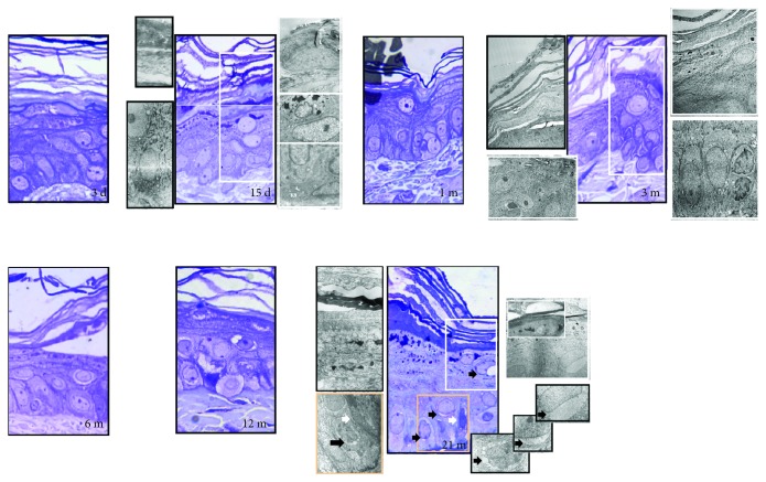
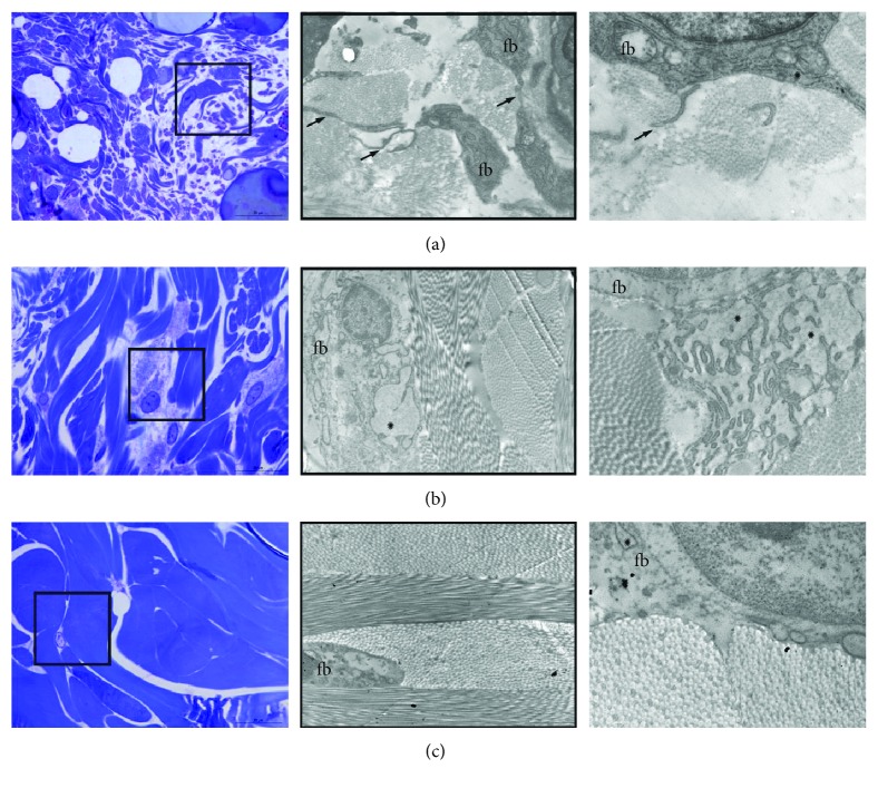
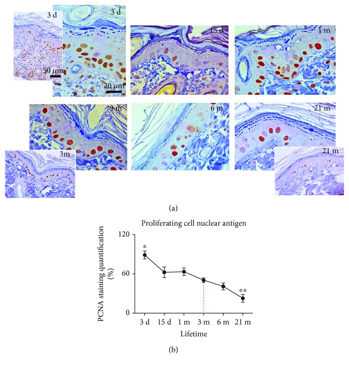
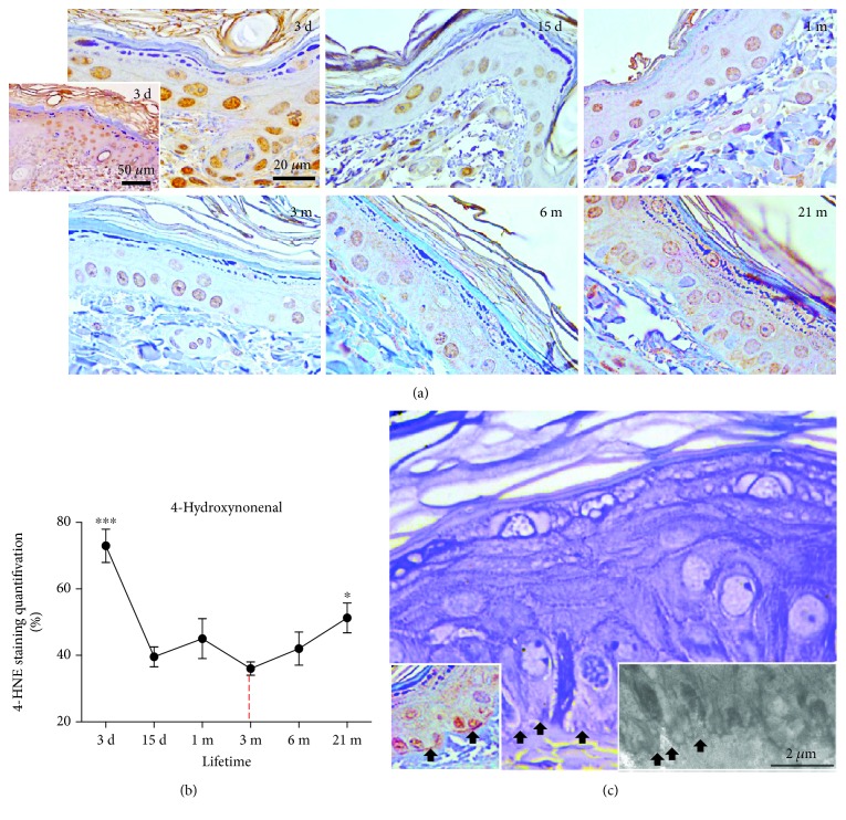
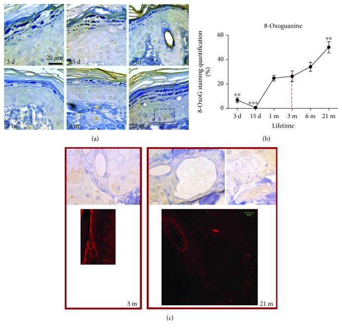
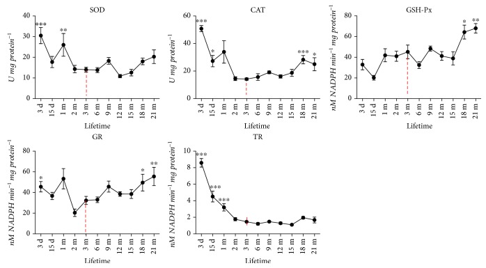
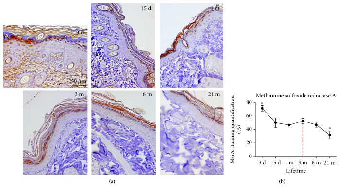
Accumulation of oxidative insults on molecular and supramolecular levels could compromise renewal potency and architecture in the aging skin. To examine and compare morphological and ultrastructural changes with redox alterations during chronological skin aging, activities of antioxidant defense (AD) enzymes, superoxide dismutase (SOD), catalase (CAT), glutathione peroxidase (GSH-Px), glutathione reductase (GR), thioredoxin reductase (TR), and methionine sulfoxide reductase A (MsrA), and the markers of oxidative damage of biomolecules-4-hydroxynonenal (HNE) and 8-oxoguanine (8-oxoG)-were examined in the rat skin during life (from 3 days to 21 months). As compared to adult 3-month-old skin, higher activities of CAT, GSH-Px, and GR and a decline in expression of MsrA are found in 21-month-old skin. These changes correspond to degenerative changes at structural and ultrastructural levels in epidermal and dermal compartments, low proliferation capacity, and higher levels of HNE-modified protein aldehydes (particularly in basal lamina) and 8-oxoG positivity in nuclei and mitochondria in the sebaceous glands and root sheath. In 3-day-old skin, higher activities of AD enzymes (SOD, CAT, GR, and TR) and MsrA expression correspond to intensive postnatal development and proliferation. In contrast to 21-month-old skin, a high level of HNE in young skin is not accompanied by 8-oxoG positivity or any morphological disturbances. Observed results indicate that increased activity of AD enzymes in elderly rat skin represents the compensatory response to accumulated oxidative damage of DNA and proteins, accompanied by attenuated repair and proliferative capacity, but in young rats the redox changes are necessary and inherent with processes which occur during postnatal skin development. Мorphological and ultrastructurаl changes are in line with the redox profile in the skin of young and old rats.


Figures

Figure 1
Figure 1
Light and electron microscopy of age-related structural changes in the rat skin. In the young skin, all epidermal cell layers have a normal structure and all cells appeared to be healthy (3 days, 15 days, 1 month, and 3 months old). Aging (6-month-, 12-month-, and 21-month-old skin) increases the epidermal layer thickness and the intercellular space throughout the epidermis along with a decrease in epithelial-dermal junction. In old skin, the basal lamina was partially detached and disrupted from the basal cell layer. Desquamation, through reduction in intercellular lipid contents, was seen in the upper layer of the stratum corneum. Intracytoplasmic vacuolization in the keratinocytes increased with aging. Namely, the keratinocyte mitochondria become light and swollen and concentrated around the nucleus to form an electron-lucent structure—“nuclear halo” (black arrow, 21 m, right insets). The numerous small cytoplasmic vacuoles, located near the nucleus, were observed in the basal keratinocytes gradually becoming a large cytoplasmic vacuole. Degenerative cells with a dark cytoplasm (white arrow, 21 m) were also seen in the basal layer of the epidermis. Semifine sections stained with toluidine blue, magnification: ×100, orig.; transmission electron microscopy, magnification: ×8000, orig.
Figure 2
Figure 2
Lifetime ultrastructural characteristics of fibroblasts and collagen. The dermis in the skin of 15-day-old rats is occupied with numerous elongated fibroblasts (a). The extracellular matrix is poorly organized, and sparse thin collagen bundles are visualized between fine fibroblast extensions (arrows). The mature skin dermis in adult 3-month-old rat (b) consisted of large fibroblasts with well-developed rough endoplasmic reticulum (ER). Dilated ER cisterns are filled with proteinaceous material; the extracellular matrix is well organized, showing compact and regularly oriented collagen bundles (b). In the dermis of 21-month-old rat, the fibroblasts reduced in number and their long cytoplasmic extensions wrap large collagen bundles with thicker and tightly packed, but poorly oriented and electron-lucent, fibers (c). Magnification: semifine section, toluidine blue staining: ×100, orig.; framed area analyzed on transmission electron microscopy, magnification: ×3000, orig.; far right: ×8800, orig.
Figure 3
Figure 3
Expression of proliferating cell nuclear antigen (PCNA) in the skin of 3-day-, 15-day-, 1-month-, 3-month-, 6-month-, and 21-month-old rats. Tissues were subjected to immunohistochemistry using a PCNA-specific antibody. The brown nuclei testify to the specific staining (a). The quantitative evaluation of PCNA antibody staining intensity in skin sections was analyzed by the IHC Profiler using the open-source ImageJ program. The relative mean intensity was determined from 6 images for each aging group (n = 6), and it is shown graphically (b). Data were presented as mean ± SEM. In respect to 3-month-old rats; p < 0.05; ∗∗p < 0.01. ×100 magnification, orig.; corner insets show low magnification (×40, orig.) of representative skin sections.
Figure 4
Figure 4
Time-course changes in the levels of HNE-modified proteins in the skin of 3-day-, 15-day-, 1-month-, 3-month-, 6-month-, and 21-month-old rats. The immunohistochemical method was used to detect skin tissue proteins modified by 4-hydroxynonenal (HNE) for subsequent quantitative image analysis (b). The brown staining testifies to the specific reaction. Strong HNE immunopositivity of the basal lamina, along with its partial discontinuation/disruption (black arrows), was observed in 21-month-old rat skin (c, left inset, arrows). Dermoepidermal junctions and the basal cell deep projections into the dermis were also disturbed (c, middle and right, arrows). (a) Representative slides of HNE immunohistochemistry are shown at ×40 (corner inset) and ×100 magnification, orig. The quantitative evaluation of HNE antibody staining intensity in skin sections (b) was analyzed as described in Figure 3. (c) Magnification: semifine section, toluidine blue staining: ×100, orig. Insets: left—HNE immunohistochemistry: ×100 orig.; right—transmission electron microscopy, bar: 2 μm. The relative mean intensity was determined from 6 images for each aging group (n = 6). Data were presented as mean ± SEM. In respect to 3-month-old rats; p < 0.05; ∗∗∗p < 0.001.
Figure 5
Figure 5
Analysis of the DNA lesion and 8-oxoguanine (8-OxoG) in the skin of rats during the lifetime. Tissues were subjected to immunohistochemistry using an 8-OxoG-specific antibody. The brown staining testifies to the specific reaction (a). The quantitative evaluation of 8-OxoG antibody staining intensity in skin sections was analyzed as described in Figure 3 (b). The relative mean intensity was determined from 6 images for each aging group (n = 6). Data were presented as mean ± SEM. In respect to 3-month-old rats; ∗∗p < 0.01; ∗∗∗p < 0.001. The mitochondrial positivity of 8-OxoG corresponds to apoptotic positivity observed by propidium iodide staining in the sebaceous glands and root sheath (c, right). Magnification: ×100, orig.
Figure 6
Figure 6
Age-dependent changes of the total superoxide dismutase (SOD), catalase (CAT), glutathione peroxidase (GSH-Px), glutathione reductase (GR), and thioredoxin reductase (TR) activities in the skin of rats. Data were presented as mean ± SEM. In respect to 3-month-old rats; p < 0.05; ∗∗p < 0.01; ∗∗∗p < 0.001.
Figure 7
Figure 7
Expression of methionine sulfoxide reductase A (MsrA) in the skin of 3-day-, 15-day-, 1-month-, 3-month-, 6-month-, and 21-month-old rats. Tissues were subjected to immunohistochemistry using an MsrA-specific antibody. The brown staining testifies to the specific reaction. The quantitative evaluation of MsrA antibody staining intensity in skin sections (b) was analyzed as described in Figure 3. The relative mean intensity was determined from 6 images for each aging group (n = 6). Data were presented as mean ± SEM.In respect to 3-month-old rats; p < 0.05. Representative slides are shown at ×40 magnification, orig.

Similar articles

Cited by

References

    1. Harman D. Aging: a theory based on free radical and radiation chemistry. Journal of Gerontology. 1956;11(3):298–300. doi: 10.1093/geronj/11.3.298. - DOI - PubMed
    1. Yoshimura H. Organ system in adaptation: the skin. In: Wilber C. G., editor. Handbook of Physiology. Washington, D.C.: American Physiological Society; 1963. pp. 109–133.
    1. Nikolakis G., Makrantonaki E., Zouboulis C. C. Skin mirrors human aging. Hormone Molecular Biology and Clinical Investigation. 2013;16(1):13–28. doi: 10.1515/hmbci-2013-0018. - DOI - PubMed
    1. Michelson A. M. Liposomal SOD and its application to skin diseases and other disorders. Japanese Journal of Dermatology. 1986;96:1376–1383.
    1. Darr D., Fridovich I. Free radicals in cutaneous biology. Journal of Investigative Dermatology. 1994;102(5):671–675. doi: 10.1111/1523-1747.ep12374036. - DOI - PubMed

MeSH terms