Review
. 2019 Mar 7;5(3):FSO373.
doi: 10.4155/fsoa-2018-0087. eCollection 2019 Mar.

Mass spectrometry-based intraoperative tumor diagnostics

Affiliations
Review

Mass spectrometry-based intraoperative tumor diagnostics

Lorena Hänel et al. Future Sci OA. .

Abstract

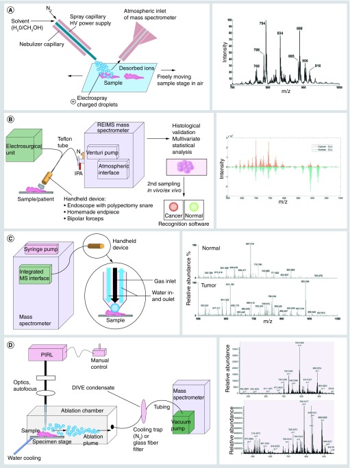
In surgical oncology, decisions regarding the amount of tissue to be removed can have important consequences: the decision between preserving sufficient healthy tissue and eliminating all tumor cells is one to be made intraoperatively. This review discusses the latest technical innovations for a more accurate tumor margin localization based on mass spectrometry. Highlighting the latest mass spectrometric inventions, real-time diagnosis seems to be within reach; focusing on the intelligent knife, desorption electrospray ionization, picosecond infrared laser and MasSpec pen, the current technical status is evaluated critically concerning its scientific and medical practice.

Keywords: DESI; MasSpec pen; PIRL; cancer; iKnife; intraoperative tumor diagnostics; real-time diagnosis; surgery; tumor margin.


Conflict of interest statement

Financial & competing interest disclosure M Kwiatkowski thanks the Forschungszentrum Medizintechnik Hamburg (FMTHH, UKE Hamburg, Germany) and the European Respiratory Society (ERS, RESPIRE3, project reference: R3201703-00121) for financial support. L Hänel was supported by the IRTG of the SFB 877 (CAU Kiel). The authors have no other relevant affiliations or financial involvement with any organization or entity with a financial interest in or financial conflict with the subject matter or materials discussed in the manuscript apart from those disclosed. No writing assistance was utilized in the production of this manuscript.

Figures

<b>Figure 1.</b>
Figure 1.. Schematic figure of mass spectrometry based methods for intraoperative cancer diagnosis (left) with corresponding examples of mass spectra (right).
(A) Desorption electrospray ionization. Left: Schematic representation of desorption electrospray ionization. Under the influence of high voltage a methanol-water solution is sprayed on the sample surface, dissolving desorbed ions to be transferred in the atmospheric inlet of the mass spectrometer. Parameters like voltage, gas and liquid flow rate can be set. Right: Average lipid profile spectrum for all pixels images used for chemical prediction. Reproduced with permission from [45]. (B) iKnife. Left: Schematic illustration of rapid evaporative mass spectrometry coupled to eligible surgical devices. Surgical ion source and ion transfer setups for rapid evaporative mass spectrometry experiments are demonstrated using an endoscope, monopolar electrosurgery or commercially available bipolar electrosurgery. The aerosol is aspirated by an air jet pump through a teflon tube with a maximum of 3 m length. By histological validation and Principal Component Analysis, in a second run the recognition software differentiates cancer and normal tissue by comparing signal intensities in the recorded mass spectra. Most of the signals in the spectra represent lipid ions. Right: Mean spectral intensity for cancer and normal tissues during cutting. Reproduced with permission from [50]. (C) MasSpec pen. Left: A handheld device (MasSpec pen) is positioned on the sample surface and through the inlet channel a water droplet is exposed for 1 s to extract molecules from the tissue surface. Then, the channel is closed and after 2 s, gas inlet is opened to transport the water droplet (volume controlled by a syringe pump) via polytetrafluorethylen (PTFE) tubing driven by vacuum into the mass spectrometer. The system is triggered by a foot pedal connected to an integrated mass spectrometer inlet. Right: Representative negative ion mode mass spectra show distinct molecular profiles from normal (average of n = 3 mass spectra) and tumor (average of n = 3 mass spectra) tissues [53]. (D) Picosecond infrared laser. Left: Current instrument for sampling of tissue with picosecond infrared laser. For cryopreserved samples, the specimen stage is cooled down to -10°C, preventing thawing during laser irradiation. The ablation chamber is sealed to capture the aerosol in its complete volume, achieved by its architecture generating a laminar flow. The aerosol is trapped through PTFE-tubing as a frozen (cooling trap) or dry (glass fiber filter) condensate within seconds. Instead of the free laser beam a fiber can be utilized to adjust and operate flexibly. Right: Negative ion mode mass spectrum from picosecond infrared laser condensate of porcine thalamus (upper) and cerebral cortex (lower) directly infused into the MS without sample preparation. HV: High voltage; IPA: Isopropyl alcohol; PIRL: Picosecond infrared laser.

Similar articles

Cited by

References

    1. Bozzetti F, Bonfanti G, Bufalino R, et al. Adequacy of margins of resection in gastrectomy for cancer. Ann. Surg. 1982;196(6):685–690. - PMC - PubMed
    1. Shin D, Park SS. Clinical importance and surgical decision-making regarding proximal resection margin for gastric cancer. World J. Gastrointest. Oncol. 2013;5(1):4–11. - PMC - PubMed
    1. Persing S, Jerome MA, James TA, et al. Surgical margin reporting in breast conserving surgery: does compliance with guidelines affect re-excision and mastectomy rates? Breast. 2015;24(5):618–622. - PMC - PubMed
    1. Heiss N, Rousson V, Ifticene-Treboux A, Lehr HA, Delaloye JF. Risk factors for positive resection margins of breast cancer tumorectomy specimen following breast-conserving surgery. Horm. Mol. Biol. Clin. Investig. 2017;32(2) - PubMed
    1. Slaughter DP, Southwick HW, Smejkal W. Field cancerization in oral stratified squamous epithelium; clinical implications of multicentric origin. Cancer. 1953;6(5):963–968. - PubMed

LinkOut - more resources