Review
. 2019 Feb;11(Suppl 2):S172-S176.
doi: 10.21037/jtd.2018.10.86.

Management of traumatic esophageal injuries

Affiliations
Review

Management of traumatic esophageal injuries

Monisha Sudarshan et al. J Thorac Dis. 2019 Feb.

Abstract

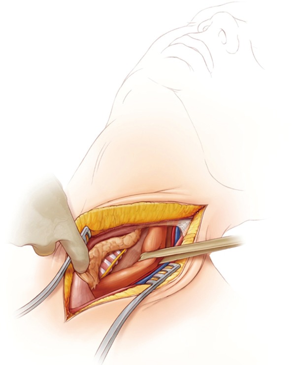
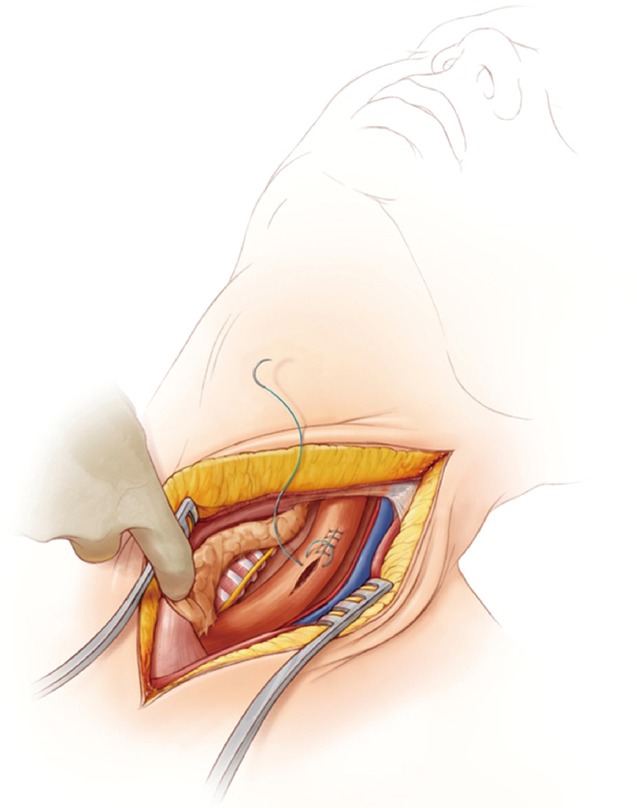
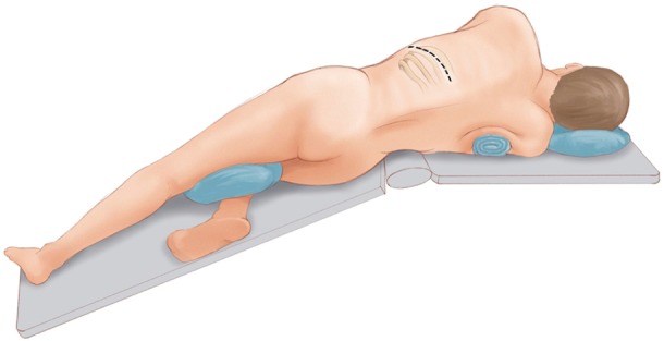
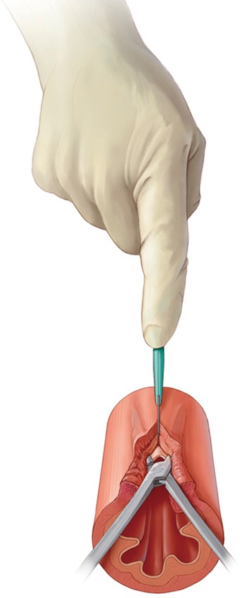
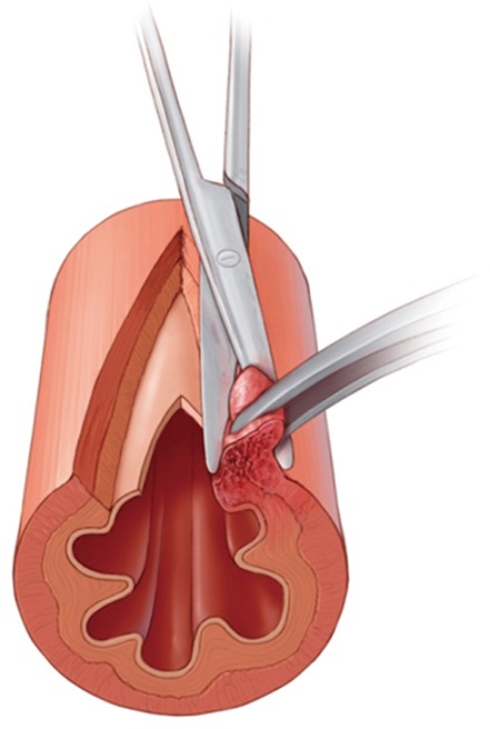
Traumatic esophageal perforations are rare but difficult to treat injuries. We review the principles of work-up, anatomy and treatment of cervical, thoracic and abdominal traumatic esophageal perforations. Management is dictated by location of the perforation and any concurrent injuries. The majority of cases are amenable to primary repair with flap re-enforcement. Other principles include adequate drainage around the repair, decompression of the esophagus and stomach (via nasogastric tube or gastrostomy tube) and distal enteral nutrition (feeding jejunostomy).

Keywords: Esophagus; esophageal leak; esophageal repair; esophageal trauma; perforations.


Conflict of interest statement

Conflicts of Interest: The authors have no conflicts of interest to declare.

Figures

Figure 1
Figure 1
Anatomy and access to the cervical esophagus.
Figure 2
Figure 2
Division of omohyoid in order to access the esophagus.
Figure 3
Figure 3
Blunt dissection of the cervical esophagus for access.
Figure 4
Figure 4
Recurrent Laryngeal Nerve in the TE groove with the esophagus encircled and retracted with a penrose drain.
Figure 5
Figure 5
Single layer closure of cervical esophageal perforation.
Figure 6
Figure 6
Patient positioning demonstrating, padding and bed flexion for left thoracotomy.
Figure 7
Figure 7
Extension of mytomy to visualize the full extent of the mucosal perforation on a thoracic esophageal perforation.
Figure 8
Figure 8
Sharp debridement of non-viable tissue on the esophagus prior to closure.
Figure 9
Figure 9
Two layer closure of thoracic esophageal injury.
Figure 10
Figure 10
Intercostal muscle flap harvested prior to entry into the left chest.
Figure 11
Figure 11
Closure of an esophageal perforation around a T tube.

References

    1. Asensio JA, Chahwan S, Forno W, et al. Penetrating esophageal injuries: multicenter study of the American Association for the Surgery of Trauma. J Trauma 2001;50:289-96. 10.1097/00005373-200102000-00015 - DOI - PubMed
    1. Patel MS, Malinoski DJ, Zhou L, et al. Penetrating oesophageal injury: a contemporary analysis of the National Trauma Data Bank. Injury 2013;44:48-55. 10.1016/j.injury.2011.11.015 - DOI - PubMed
    1. Biffl WL, Moore EE, Feliciano DV, et al. Western Trauma Association Critical Decisions in Trauma: Diagnosis and management of esophageal injuries. J Trauma Acute Care Surg 2015;79:1089-95. 10.1097/TA.0000000000000772 - DOI - PubMed
    1. Sperry JL, Moore EE, Coimbra R, et al. Western Trauma Association critical decisions in trauma: penetrating neck trauma. J Trauma Acute Care Surg 2013;75:936-40. 10.1097/TA.0b013e31829e20e3 - DOI - PubMed
    1. Makhani M, Midani D, Goldberg A, et al. Pathogenesis and outcomes of traumatic injuries of the esophagus. Dis Esophagus 2014;27:630-6. 10.1111/dote.12132 - DOI - PMC - PubMed