Review
. 2019 Jan 22;8(4):e1554969.
doi: 10.1080/2162402X.2018.1554969. eCollection 2019.

Making cold malignant pleural effusions hot: driving novel immunotherapies

Affiliations
Review

Making cold malignant pleural effusions hot: driving novel immunotherapies

Pranav Murthy et al. Oncoimmunology. .

Abstract

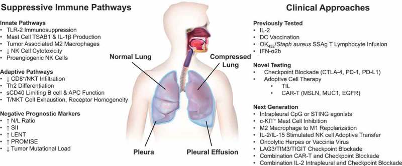
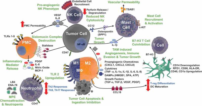
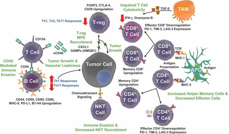
Malignant pleural effusions, arising from either primary mesotheliomas or secondary malignancies, heralds advanced disease and poor prognosis. Current treatments, including therapeutic thoracentesis and tube thoracostomy, are largely palliative. The immunosuppressive environment within the pleural cavity includes myeloid derived suppressor cells, T-regulatory cells, and dysfunctional T cells. The advent of effective immunotherapy with checkpoint inhibitors and adoptive cell therapies for lung cancer and other malignancies suggests a renewed examination of local and systemic therapies for this malady. Prior strategies reporting remarkable success, including instillation of the cytokine interleukin-2, perhaps coupled with checkpoint inhibitors, should be further evaluated in the modern era.

Keywords: Malignant pleural effusion (MPE); adoptive cell therapy (ACT); cancer immunotherapies; damage associated molecular pattern molecules (DAMPs); interleukin-2 (IL-2); mesothelioma (MM); non-small cell lung cancer (NSCLC).


Figures

Figure 1.
Figure 1.
Immunotherapy for patients with malignant pleural effusions. Malignant pleural effusions are an inflammatory condition within the chest containing immunologically active but most often exhausted cells associated with both bulk tumor and tumor cells in suspension. Thus, they are a functionally ‘cold’ site. Several suppressive innate and adaptive pathways have been identified. MPE pathophysiology is closely correlated with the upregulation of these inflammatory pathways. Systemic measures associated with acute inflammation (neutrophils) and chronic inflammation – immunity (lymphocytes) can be utilized as prognostic indicators of disease outcome and for disease stratification. Previously tested clinical approaches, including IL-2 therapy and DC vaccination hold much promise, and should be further investigated in the context of trials evaluating checkpoint inhibition and adoptive cell therapy. Next generation immunotherapies may enable personalized treatments, leverage improved cell-based therapies, and modulate the local suppressive tumor microenvironment. Local therapies could be more effectively deployed.
Figure 2.
Figure 2.
Innate immune signaling pathways in malignant pleural effusions. The altered tumor microenvironment enables MPE formation by enhancing angiogenesis, promoting vascular permeability, driving tumor growth, unscheduled cell death, and releasing damage associated molecular pattern molecules (DAMPs). Natural Killer (NK) cells exhibit a proangiogenic phenotype, with reduced cytotoxicity, increasing endothelial cell recruitment with production of IL-8 and vascular endothelial growth factor (VEGF). Pleural Mesothelial Cells (PMCs) respond to pleural cavity DAMPs and other ligands via toll-like receptor (TLR) 1–9 signaling, promoting recruitment of inflammatory cells with release of platelet derived growth factor (PDGF), IL-8, nitric oxide, and monocyte chemotactic protein 1 (MCP-1). Tumor cell production of VEGF and sialidase promote vascular permeability with destruction of the protective extracellular PMC sialomucin complex. Leukotriene B4 (LB4) and epithelial neutrophil-activating peptide-78 (ENA-78) increase neutrophil recruitment to the pleural space and neutropenia is sustained with CD47 mediated inhibition of neutrophil apoptosis. Accompanying neutrophil and macrophage (MΦ) TLR2 upregulation support MPE formation with a skewed Th2 response. Tumor associated macrophages (TAMs), favoring M2 polarization protect tumor cells from apoptosis and promote angiogenesis, immune evasion, and tumor growth with a milieu of proangiogenic chemokines, cytokines, DAMPs, and growth factors. TAMs also produce chemokine ligand 18 (CCL18), which promotes T regulatory cell (T-reg) differentiation and limits dendritic cell (DC) maturation. DCs facilitate immunosuppression via B7-H3 T cell coinhibition and upregulation of RFD1, CD86, HLA-DR, CD40, and CD1a expression. Tumor cells avoid ingestion by expressing CD47, a ‘don’t eat me’ signal and produce CC12 and osteopontin, inducing mast cell recruitment and c-KIT activation respectively. Increased mast cells are found to be associated with MPE formation with tumor growth promoting IL-1β and tryptase alpha/beta-1 (TSAB1) mediated enhanced vascular permeability.
Figure 3.
Figure 3.
Adaptive immune signaling pathways in malignant pleural effusions. The immunosuppressive MPE environment modulates the ‘adaptome’, B, NKT, αβ T cell and γδ T lymphocyte biology to promote tumor growth and immune evasion. Tumor cells secrete chemokine ligand 1 (CXCL1) and DAMPs, promoting further cell proliferation and T-regulatory (T-reg) cell recruitment. T-regs promote an immunosuppressive environment, inhibiting Th1, Th2, Th9, and Th17 responses. Moreover, T lymphocytes display a high CD4+/CD8+ ratio with an increased percentage of central memory CD4+ T cells and decreased CD8+ effector-memory T cells. T cell programmed cell death protein 1 (PD-1), T cell immunoglobulin and mucin domain 3 (TIM-3), and Lymphocyte-activation gene 3 (LAG-3) immune checkpoint expression inhibit lymphocyte activity. Tumor Associated Macrophage (TAM) transforming growth factor beta (TGF-β) production decreases effector T cell cytotoxicity with reduced production of interferon gamma (IFN-γ) and granzyme B. These changes are accompanied with decreased NKT recruitment to the effusion site. Increased soluble CD40 (sCD40) levels inhibit B cell function by competing for CD154 (CD40 ligand) on T lymphocytes. Despite decreased B cell density, increased expression of CD80, CD86, MHC II, CD44, CD69, and programmed death ligand 1 (PD-L1) promote Th2 responses that can also support MPE formation.

References

    1. Muduly D, Deo S, Subi T, Kallianpur A, Shukla N.. An update in the management of malignant pleural effusion. Indian J Palliat Care. 2011;17(2):98–103. doi: 10.4103/0973-1075.84529. - DOI - PMC - PubMed
    1. Psallidas I, Kalomenidis I, Porcel JM, Robinson BW, Stathopoulos GT.. Malignant pleural effusion: from bench to bedside. Eur Respir Rev. 2016;25(140):189–198. doi: 10.1183/16000617.0019-2016. - DOI - PMC - PubMed
    1. Zamboni MM, Da Silva CT Jr., Baretta R, Cunha ET, Cardoso GP. Important prognostic factors for survival in patients with malignant pleural effusion. BMC Pulm Med. 2015;15:29. doi: 10.1186/s12890-015-0025-z. - DOI - PMC - PubMed
    1. Lee YC. Hunting for a pleural fluid test for mesothelioma: is soluble mesothelin the answer? Thorax. 2007;62(7):561–562. doi: 10.1136/thx.2006.076612. - DOI - PMC - PubMed
    1. Cheah HM, Lansley SM, Varano Della Vergiliana JF, Tan AL, Thomas R, Leong SL, Creaney J, Lee YC. Malignant pleural fluid from mesothelioma has potent biological activities. Respirology. 2017;22(1):192–199. doi: 10.1111/resp.12874. - DOI - PubMed

Publication types

LinkOut - more resources