Review
. 2019 Feb 18:2019:6715257.
doi: 10.1155/2019/6715257. eCollection 2019.

Female Urethroplasty: A Practical Guide Emphasizing Diagnosis and Surgical Treatment of Female Urethral Stricture Disease

Affiliations
Review

Female Urethroplasty: A Practical Guide Emphasizing Diagnosis and Surgical Treatment of Female Urethral Stricture Disease

Marjan Waterloos et al. Biomed Res Int. .

Abstract

Female urethral strictures are rare. Guidelines on how to diagnose and treat these strictures are lacking. At present, only expert opinion is available to guide clinical practice. Once the diagnosis is suspected based on obstructive voiding symptoms and uroflowmetry, most clinicians will use in addition video-urodynamics (including urethrography), urethral calibration and cystourethroscopy for confirmation of the diagnosis. Clinical inspection and gynaecological examination are also important. Urethral dilation is usually the first-line treatment despite the lack of long-term success. Female urethroplasty is associated with higher success rates. A multitude of techniques are described but not one technique has shown superiority above another. This narrative review aims to provide a clinical guide for diagnosis and treatment to the urologist motivated to perform female urethroplasty.


Figures

Figure 1
Figure 1
Female versus male urethral anatomy. (a) = female urethral anatomy; (b) = male urethral anatomy. Green = ventral urethra; red = dorsal urethra.
Figure 2
Figure 2
Wine glass image. Voiding cystourethrography with a distal female urethral stricture and prestenotic dilation.
Figure 3
Figure 3
MRI of the female pelvis. The urethra is clearly visible without the presence of a urethral diverticulum or periurethral abscess.
Figure 4
Figure 4
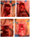
Exposure of the female urethra and vagina using the lone-star retractor.
Figure 5
Figure 5
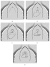
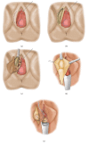
Anterior vaginal wall flap (“Blandy flap”). (a) = inverted U-incision; (b) = ventral stricturotomy; (c) & (d) = suturing the tip of the U-flap to the proximal part of the opened urethra; (e) = further suturing the edges of the flap to the urethral edges.
Figure 6
Figure 6
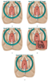
Vestibular flap (“Montorsi flap”). (a) = inverted-Y incision; (b) = dorsal stricturotomy; (c) = mobilization of the vestibular flap; (d) = suturing the tip of the flap to the proximal end of the opened urethra and the edges of the flap to the urethral edges; (e) = suturing the base of the flap to the vestibular mucosa.
Figure 7
Figure 7
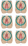
Lateral vaginal wall flap (“Orandi flap”). (a) = longitudinal midline incision at anterior vaginal wall; (b) = ventral stricturotomy; (c) = mobilization of the lateral vaginal wall flap; (d) = suturing the medial surface of the flap towards the ipsilateral side of the urethra; (e) = turning the mucosal surface of the flap towards the urethral lumen and suturing the lateral side of the flap to the contralateral side of the urethra.
Figure 8
Figure 8
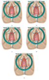
Dorsal onlay free graft urethroplasty. (a) = semilunar suprameatal incision; (b) = dorsal stricturotomy; (c) & (d) = suturing the edges of the graft to the urethral edges; (e) = suturing the distal edges of the graft to the edge of the suprameatal incision.
Figure 9
Figure 9
Dorsal onlay buccal mucosa graft urethroplasty. (a) = semilunar dorsal incision and stricturotomy with stay sutures placed at the opened urethra; (b) = quilting the graft against the surface of the clitoral bodies and suturing the edges of the graft against the urethral edges; (c) = suturing the distal edges of the graft to the edge of the suprameatal incision; (d) = final result.
Figure 10
Figure 10
Ventral onlay free graft urethroplasty. (a) = semilunar inframeatal incision; (b) = ventral stricturotomy; (c and d) = suturing the edges of the graft to the urethral edges; (e) = suturing the distal edges of the graft to the edge of the inframeatal incision.
Figure 11
Figure 11
Martius flap. (a) & (b) = sagittal incision at the most dependent line of the labium majus; (c) & (d) = mobilizing fibrofatty tissue; (e) = transposition of the Martius flap to the reconstructed area through a subcutaneous tunnel.
Figure 12
Figure 12
Martius flap procedure after ventral urethral repair. (a) = sagittal incision line; (b) = lateral dissection to the labiocrural fold; (c) = medial dissection up to the bulbospongiosus muscle; (d) = mobilization of the flap; (e) = division of the anterior pedicle; (f) = transposed Martius flap to the ventrally reconstructed area.

References

    1. Ackerman A. L., Blaivas J., Anger J. T. Female urethral reconstruction. Current Bladder Dysfunction Reports. 2010;5(4):225–232. doi: 10.1007/s11884-010-0071-6. - DOI - PMC - PubMed
    1. Faiena I., Koprowski C., Tunuguntla H. Female urethral reconstruction. The Journal of Urology. 2016;195(3):557–567. doi: 10.1016/j.juro.2015.07.124. - DOI - PubMed
    1. Hoag N., Chee J. Surgical management of female urethral strictures. Translational Andrology and Urology. 2017;6(Suppl 2):S76–S80. doi: 10.21037/tau.2017.01.20. - DOI - PMC - PubMed
    1. Osman N. I., Mangera A., Chapple C. R. A systematic review of surgical techniques used in the treatment of female urethral stricture. European Urology. 2013;64(6):965–973. doi: 10.1016/j.eururo.2013.07.038. - DOI - PubMed
    1. Lumen N., Hoebeke P., Oosterlinck W. Urethroplasty for urethral strictures: quality assessment of an in-home algorithm. International Journal of Urology. 2010;17(2):167–174. doi: 10.1111/j.1442-2042.2009.02435.x. - DOI - PubMed

LinkOut - more resources