. 2019 Feb 13:2019:8520856.
doi: 10.1155/2019/8520856. eCollection 2019.

Inhibition of HDAC3 Ameliorates Cerebral Ischemia Reperfusion Injury in Diabetic Mice In Vivo and In Vitro

Affiliations

Inhibition of HDAC3 Ameliorates Cerebral Ischemia Reperfusion Injury in Diabetic Mice In Vivo and In Vitro

Bo Zhao et al. J Diabetes Res. .

Abstract

Background: A substantial increase in histone deacetylase 3 (HDAC3) expression is implicated in the pathological process of diabetes and stroke. However, it is unclear whether HDAC3 plays an important role in diabetes complicated with stroke. We aimed to explore the role and the potential mechanisms of HDAC3 in cerebral ischemia/reperfusion (I/R) injury in diabetic state.

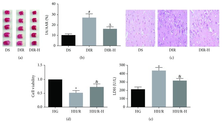
Methods: Diabetic mice were subjected to 1 h ischemia, followed by 24 h reperfusion. PC12 cells were exposed to high glucose for 24 h, followed by 3 h of hypoxia and 6 h of reoxygenation (H/R). Diabetic mice received RGFP966 (the specific HDAC3 inhibitor) or vehicle 30 minutes before the middle cerebral artery occlusion (MCAO), and high glucose-incubated PC12 cells were pretreated with RGFP966 or vehicle 6 h before H/R.

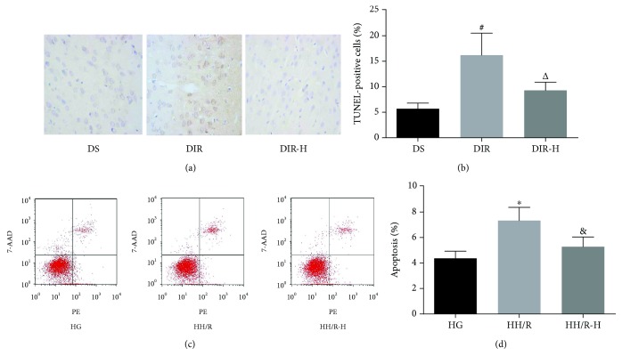
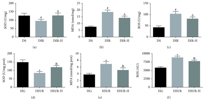
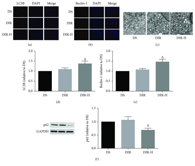
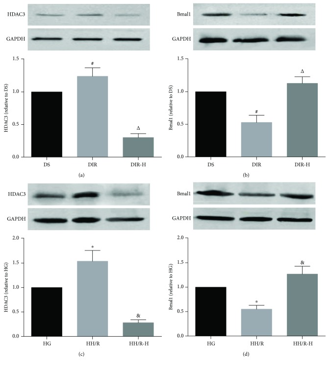
Results: HDAC3 inhibition reduced the cerebral infarct volume, ameliorated pathological changes, improved the cell viability and cytotoxicity, alleviated apoptosis, attenuated oxidative stress, and enhanced autophagy in cerebral I/R injury model in diabetic state in vivo and in vitro. Furthermore, we found that the expression of HDAC3 was remarkably amplified, and the Bmal1 expression was notably decreased in diabetic mice with cerebral I/R, whereas this phenomenon was obviously reversed by RGFP966 pretreatment.

Conclusions: These results suggested that the HDAC3 was involved in the pathological process of the complex disease of diabetic stroke. Suppression of HDAC3 exerted protective effects against cerebral I/R injury in diabetic state in vivo and in vitro via the modulation of oxidative stress, apoptosis, and autophagy, which might be mediated by the upregulation of Bmal1.


Figures

Figure 1
Figure 1
Inhibition of HDAC3 played protective effects against cerebral I/R injury. Brain tissues were collected for determination of cerebral infarct volume by TTC staining (a, b) and observation of pathological changes by H&E staining (c) in each group. PC12 cells were collected for assessment of cell viability assessed by CCK-8 (d) and cytotoxicity by lactate dehydrogenase (LDH) release (e) in each group. All values are presented as mean ± SD, n = 6/group. #P < 0.05 versus the DS group, P < 0.05 versus the DIR group, P < 0.05 versus the HG group, and &P < 0.05 versus the HH/R group.
Figure 2
Figure 2
Inhibition of HDAC3 alleviated I/R-induced apoptosis. Apoptosis in the brain tissues was assessed by TUNEL staining (a, b); in the PC12 cells, it was evaluated by flow cytometry (c, d) in each group. All values are presented as mean ± SD, n = 6/group. #P < 0.05 versus the DS group, P < 0.05 versus the DIR group, P < 0.05 versus the HG group, and &P < 0.05 versus the HH/R group.
Figure 3
Figure 3
Inhibition of HDAC3 attenuated I/R-induced oxidative stress. Brain tissues were collected for analysis of SOD activity (a), MDA content (b), and ROS levels (c); PC12 cells were also collected for detection of SOD activity (d), MDA content (e), and ROS levels (f). All values are presented as mean ± SD, n = 6/group. #P < 0.05 versus the DS group, P < 0.05 versus the DIR group, P < 0.05 versus the HG group, and &P < 0.05 versus the HH/R group.
Figure 4
Figure 4
HDAC3 inhibition enhanced autophagy in diabetic mice subjected to cerebral I/R injury. Representative immunofluorescence staining images and assessment of LC3B (a, d) and beclin-1 (b, e) (×100). Representative electron micrographs of each group (c) (×5000). Representative Western blot images and assessment of p62 (f) level in each group. All results are presented as mean ± SD, n = 6/group. #P < 0.05 versus the DS group and P < 0.05 versus the DIR group.
Figure 5
Figure 5
The protective effects of RGFP966 were mediated by the upregulation of Bmal1. Representative Western blot images and assessment of the HDAC3 and Bmal1 level in each group. HDAC3 and Bmal1 expression in the mouse brain (a, b) and HDAC3 and Bmal1 expression in PC12 cells (c, d). All results are presented as mean ± SD, n = 6/group. #P < 0.05 versus the DS group, P < 0.05 versus the DIR group, P < 0.05 versus the HG group, and &P < 0.05 versus the HH/R group.

Similar articles

Cited by

References

    1. Liu L., Wang D., Wong K. S. L., Wang Y. Stroke and stroke care in China: huge burden, significant workload, and a national priority. Stroke. 2011;42(12):3651–3654. doi: 10.1161/STROKEAHA.111.635755. - DOI - PubMed
    1. Schellinger P. D., Kohrmann M. 4.5-hour time window for intravenous thrombolysis with recombinant tissue-type plasminogen activator is established firmly. Stroke. 2014;45(3):912–913. doi: 10.1161/STROKEAHA.113.002700. - DOI - PubMed
    1. Shou J., Zhou L., Zhu S., Zhang X. Diabetes is an independent risk factor for stroke recurrence in stroke patients: a meta-analysis. Journal of Stroke & Cerebrovascular Diseases. 2015;24(9):1961–1968. doi: 10.1016/j.jstrokecerebrovasdis.2015.04.004. - DOI - PubMed
    1. Osei E., den Hertog H. M., Berkhemer O. A., et al. Admission glucose and effect of intra-arterial treatment in patients with acute ischemic stroke. Stroke. 2017;48(5):1299–1305. doi: 10.1161/STROKEAHA.116.016071. - DOI - PubMed
    1. Pan Y., Jing J., Li H., Wang Y., Wang Y., He Y. Abnormal glucose regulation increases stroke risk in minor ischemic stroke or TIA. Neurology. 2016;87(15):1551–1556. doi: 10.1212/WNL.0000000000003200. - DOI - PubMed

MeSH terms

LinkOut - more resources