. 2019 Sep 2:4:29.
doi: 10.12688/wellcomeopenres.15066.2. eCollection 2019.

Spo13 prevents premature cohesin cleavage during meiosis

Affiliations

Spo13 prevents premature cohesin cleavage during meiosis

Stefan Galander et al. Wellcome Open Res. .

Abstract

Background: Meiosis produces gametes through two successive nuclear divisions, meiosis I and meiosis II. In contrast to mitosis and meiosis II, where sister chromatids are segregated, during meiosis I, homologous chromosomes are segregated. This requires the monopolar attachment of sister kinetochores and the loss of cohesion from chromosome arms, but not centromeres, during meiosis I. The establishment of both sister kinetochore mono-orientation and cohesion protection rely on the budding yeast meiosis I-specific Spo13 protein, the functional homolog of fission yeast Moa1 and mouse MEIKIN. Methods: Here we investigate the effects of loss of SPO13 on cohesion during meiosis I using a live-cell imaging approach. Results: Unlike wild type, cells lacking SPO13 fail to maintain the meiosis-specific cohesin subunit, Rec8, at centromeres and segregate sister chromatids to opposite poles during anaphase I. We show that the cohesin-destabilizing factor, Wpl1, is not primarily responsible for the loss of cohesion during meiosis I. Instead, premature loss of centromeric cohesin during anaphase I in spo13 Δ cells relies on separase-dependent cohesin cleavage. Further, cohesin loss in spo13 Δ anaphase I cells is blocked by forcibly tethering the regulatory subunit of protein phosphatase 2A, Rts1, to Rec8. Conclusions: Our findings indicate that separase-dependent cleavage of phosphorylated Rec8 causes premature cohesin loss in spo13 Δ cells.

Keywords: Meiosis; Spo13; cohesin.


Conflict of interest statement

No competing interests were disclosed.

Figures

Figure 1.
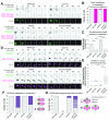
Figure 1.. Cohesin is lost at anaphase I in the absence of SPO13.
( A) Representative images of Rec8-GFP, Mtw1-tdTomato and Pds1-tdTomato in live sporulating wild-type (AM13716), spo13Δ (AM15133), mam1Δ (AM15134) and spo13Δ mam1Δ (AM15135) cells. Scale bars represent 1 µm. Arrows indicate pericentromeric cohesin. ( B) The number of cells with pericentromeric Rec8-GFP in anaphase I is shown after scoring 50 cells from ( A). ( C) Rec8-GFP intensity was measured for 50 cells from ( A) in the area occupied by the tdTomato-labeled kinetochore protein Mtw1. ***p<0.001 (Welch two-sample t-test). ( D) Rec8 loading is unaffected by deletion of SPO13. Rec8-3Ha association with the indicated sites was measured in prophase I in wild-type (AM4015), spo13Δ (AM15343), mam1Δ (AM15342) and spo13Δ mam1Δ (AM15344) cells carrying ndt80Δ and a no tag control (AM11633). Cells were arrested in prophase by harvesting 5 h after resuspension in sporulation medium and anti-Ha ChIP-qPCR performed. Error bars show standard error of the mean from three independent biological experiments.
Figure 2.
Figure 2.. Deletion of SPO13 permits sister chromosome segregation in anaphase I in mam1Δ mutants.
( A) Assay for mono-orientation and cohesion defects using heterozygous centromeric fluorescent markers. Representative images are shown. Scale bars represent 1 µm. Images for Δ CEN5=0µm, Δ CEN5=0-2µm and Δ CEN5>2µm were taken from wild-type, mam1Δ and spo13Δ cells, respectively. ( B) Frequency of CEN5 distance categories is shown for the indicated genotypes after live-cell imaging. Wild-type (AM15190), spo13Δ (AM15118), mam1Δ (AM15119) and spo13Δ mam1Δ (AM15120) cells carrying SPC42-tdTomato, PDS1-tdTomato and heterozygous TetR-GFP foci at CEN5, were sporulated for 2.5 h before imaging on a microfluidics plate.
Figure 3.
Figure 3.. Deletion of MAD2 restores the second nuclear division, but not accurate chromosome segregation to spo13Δ mutants.
( AC) Representative images of Rec8-GFP, Mtw1-tdTomato and Pds1-tdTomato in live sporulating wild-type (AM13716), spo13Δ (AM24843), mad2Δ (AM24844) and spo13Δ mad2Δ (AM24845) cells. Scale bars represent 1 µm. Arrows indicate pericentromeric cohesin. ( B) The number of cells with pericentromeric Rec8-GFP in anaphase I is shown after scoring 50 cells from ( A). ( C) Rec8-GFP intensity was measured for 50 cells from ( A) as described for Figure 1C. ***p<0.001, n.s. = not significant (Welch two-sample t-test). For spo13Δ mad2Δ mutants, the analysis in ( B) and ( C) was performed exclusively for cells that performed two divisions (as judged by the presence of four Mtw1-tdTomato foci after meiosis II). ( DG) Severe chromosome missegregation occurs in spo13Δ mad2Δ cells. ( D) Representative images of wild-type (AM24848), spo13Δ (AM24849), mad2Δ (AM25221) and spo13Δ mad2Δ (AM25222) cells carrying heterozygous TetR-GFP foci at CEN5 and HTB1-mCherry. Green arrows indicate CEN5-GFP segregation outcomes after meiosis I, cyan arrows indicate CEN5-GFP segregation outcomes after meiosis II. ( E) Spores of spo13Δ mad2Δ vary greatly in the amount of nuclear DNA, as estimated by Htb1-mCherry area, thus indicating gross chromosome missegregation. The area occupied by Htb1-mCherry was measured in cells with four (wild type (n=45), mad2Δ (n=31) and spo13Δ mad2Δ (n=33)), or two ( spo13Δ (n=50)) nuclear masses after meiosis II and variation in chromosomal area estimated by obtaining the ratio of the largest and smallest nuclear mass for each cell. **p<0.01, n.s. = not significant (Welch two-sample t-test). ( FG) CEN5 missegregation in spo13Δ mad2Δ cells. Segregation of heterozygous CEN5-GFP foci was scored in 50 cells after the first ( F) and second ( G) nuclear division in the indicated strains. For spo13Δ mad2Δ mutants, the analysis in ( F) and ( G) was performed exclusively for cells that performed two divisions (as judged by the presence of four distinct Htb1-mCherry signals after meiosis II). Note that while a large proportion of spo13Δ mad2Δ cells end up with CEN5-GFP foci in two separate spores after meiosis II (similar to wild type), many of these cells have already segregated sister chromosomes in meiosis I (purple stripes), rather than meiosis II (gray).
Figure 4.
Figure 4.. Cohesin protection in spo13Δ cells is rescued by inhibition of separase, but not by ablation of the prophase pathway.
( A) Deletion of RAD61/WPL1 does not rescue sister chromatid cohesion in spo13Δ cells. Categorization of CEN5-GFP distances in wild-type (AM15190), spo13Δ (AM20146), rad61Δ (AM21068) and spo13Δ rad61Δ (AM21358) cells carrying SPC42-tdTomato, PDS1-tdTomato and heterozygous TetR-GFP dots at CEN5 was carried out as described in Figure 2A. ( BD) Separase activity is required for Rec8 removal in spo13Δ mutants. Wild-type (AM13716), spo13Δ (AM20033), esp1-2 (AM20868) and spo13Δ esp1-2 (AM21949) cells carrying REC8-GFP, MTW1-tdTomato and PDS1-tdTomato were resuspended in sporulation medium at 32°C and grown in flasks for 3h before transferring to a microfluidics plate and imaged at 32°C. ( B) The number of cells with the indicated patterns of Rec8-GFP localization in anaphase I was scored for 50 cells per strain. ( C) The intensity of pericentromeric Rec8-GFP for the indicated genotypes is shown. The mean of the two maximum intensity values on a straight line connecting both kinetochores in anaphase I (within the first two time points after Pds1-tdTomato degradation) was measured for 50 cells. Error bars represent standard error. ***p<0.001 (Welch two-sample t-test). ( D) Representative images are shown. Scale bars represent 1 µm. Arrows indicate pericentromeric cohesin. ( E) Inhibition of separase activity restores sister chromatid cohesion to spo13Δ mutants. Cohesion was assayed by categorization of CEN5-GFP distances as described in Figure 2A. Strains used were wild-type (AM15190), spo13Δ (AM20146), esp1-2 (AM22498) and spo13Δ esp1-2 (AM22499) cells carrying SPC42-tdTomato, PDS1-tdTomato and heterozygous TetR-GFP dots at CEN5.
Figure 5.
Figure 5.. Cohesin cleavage is required for loss of sister chromatid cohesion in spo13Δ cells.
( AC) Non-cleavable Rec8 blocks efficient removal of cohesin in spo13Δ cells. ( A) Representative images from movies of cells carrying Rec8-GFP, Mtw1-tdTomato, Pds1-tdTomato and with the indicated genotypes are shown. Scale bars represent 1 µm. Arrows indicate pericentromeric Rec8-GFP. ( B) Frequency of cells with the indicated pattern of Rec8-GFP localization is shown for the indicated genotypes. ( C) Rec8-GFP intensity was measured for the indicated genotypes as described in Figure 4C. Error bars represent standard error. **p<0.01, ***p<0.001 (Welch two-sample t-test). Strains used in ( AC) were REC8-GFP (AM22190), REC8-GFP spo13Δ (AM22191), rec8-N-GFP (AM22192) and rec8-N-GFP spo13Δ (AM22193) cells carrying MTW1-tdTomato and PDS1-tdTomato. ( D) Non-cleavable Rec8 prevents sister chromatid segregation in spo13Δ mutants. Cohesion functionality was determined for the indicated genotypes by categorization of CEN5-GFP distances as described for Figure 2A. Strains were REC8-3HA (AM22346), REC8-3HA spo13Δ (AM22347), rec8-N-3HA (AM22348) and rec8-N-3HA spo13Δ (AM22349) and carried SPC42-tdTomato, PDS1-tdTomato and heterozygous TetR-GFP dots at CEN5.
Figure 6.
Figure 6.. PP2A can prevent cohesin cleavage and sister chromatid segregation in spo13Δ cells.
( AC) Cohesin is retained on chromosomes when PP2A Rts1 is tethered to Rec8. ( A) Representative images of Rec8-GFP, Mtw1-dtTomato and Pds1-tdTomato in wild-type (AM13716), spo13Δ (AM20033), pCLB2-SGO1 (AM21315), RTS1-GBP (AM21316), spo13Δ pCLB2-SGO1 (AM21317), spo13Δ RTS1-GBP (AM21319), pCLB2-SGO1 RTS1-GBP (AM21318) and spo13Δ pCLB2-SGO1 RTS1-GBP (AM21320) cells undergoing meiosis. Scale bars represent 1 µm. Arrows indicate pericentromeric cohesin. ( B) The number of cells with pericentromeric cohesin in anaphase I was scored for 50 cells per strain. ( C) Rec8-GFP intensity in anaphase I was measured as described in Figure 2A. **p<0.01, ***p<0.001 (Welch two-sample t-test).
Figure 7.
Figure 7.. Fusion of Rts1 to a separase biosensor prevents its cleavage in both wild-type and spo13Δ cells.
( A) Schematic illustration of the separase biosensor and its Rts1 fusion. ( B and C) Wild-type (AM21557) and spo13Δ cells (AM21558) carrying a wild-type separase biosensor ( pCUP1-GFP-REC8(110-500)-LacI) or an Rts1 fused biosensor ( pCUP1-GFP-REC8(110-500)-LacI-RTS1; wild type: AM21559, spo13Δ: AM21800) as well as lys2::lacOx256 and PDS1-tdTomato were sporulated in the presence of 100 nM CuSO 4 for 2.5 h before imaging on a microfluidics plate. ( B) Representative images are shown. Scale bars represent 1 µm. ( C) Scoring of 50 cells per strain for the presence of GFP foci (uncleaved biosensor) or diffuse GFP signal (cleaved biosensor) within 30 min (two time points) of Pds1 degradation.

References

    1. Argüello-Miranda O, Zagoriy I, Mengoli V, et al. : Casein Kinase 1 Coordinates Cohesin Cleavage, Gametogenesis, and Exit from M Phase in Meiosis II. Dev Cell. 2017;40(1):37–52. 10.1016/j.devcel.2016.11.021 - DOI - PubMed
    1. Attner MA, Miller MP, Ee LS, et al. : Polo kinase Cdc5 is a central regulator of meiosis I. Proc Natl Acad Sci U S A. 2013;110(35):14278–14283. 10.1073/pnas.1311845110 - DOI - PMC - PubMed
    1. Brar GA, Kiburz BM, Zhang Y, et al. : Rec8 phosphorylation and recombination promote the step-wise loss of cohesins in meiosis. Nature. 2006;441(7092):532–536. 10.1038/nature04794 - DOI - PubMed
    1. Buheitel J, Stemmann O: Prophase pathway-dependent removal of cohesin from human chromosomes requires opening of the Smc3-Scc1 gate. EMBO J. 2013;32(5):666–676. 10.1038/emboj.2013.7 - DOI - PMC - PubMed
    1. Buonomo SB, Clyne RK, Fuchs J, et al. : Disjunction of homologous chromosomes in meiosis I depends on proteolytic cleavage of the meiotic cohesin Rec8 by separin. Cell. 2000;103(3):387–398. 10.1016/S0092-8674(00)00131-8 - DOI - PubMed

LinkOut - more resources