Probing High Permeability of Nuclear Pore Complexes by Scanning Electrochemical Microscopy: Ca2+ Effects on Transport Barriers
- PMID: 30907572
- PMCID: PMC6535230
- DOI: 10.1021/acs.analchem.9b00796
Probing High Permeability of Nuclear Pore Complexes by Scanning Electrochemical Microscopy: Ca2+ Effects on Transport Barriers
Abstract
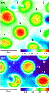
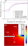
The nuclear pore complex (NPC) solely mediates molecular transport between the nucleus and cytoplasm of a eukaryotic cell to play important biological and biomedical roles. However, it is not well-understood chemically how this biological nanopore selectively and efficiently transports various substances, including small molecules, proteins, and RNAs by using transport barriers that are rich in highly disordered repeats of hydrophobic phenylalanine and glycine intermingled with charged amino acids. Herein, we employ scanning electrochemical microscopy to image and measure the high permeability of NPCs to small redox molecules. The effective medium theory demonstrates that the measured permeability is controlled by diffusional translocation of probe molecules through water-filled nanopores without steric or electrostatic hindrance from hydrophobic or charged regions of transport barriers, respectively. However, the permeability of NPCs is reduced by a low millimolar concentration of Ca2+, which can interact with anionic regions of transport barriers to alter their spatial distributions within the nanopore. We employ atomic force microscopy to confirm that transport barriers of NPCs are dominantly recessed (∼80%) or entangled (∼20%) at the high Ca2+ level in contrast to authentic populations of entangled (∼50%), recessed (∼25%), and "plugged" (∼25%) conformations at a physiological Ca2+ level of submicromolar. We propose a model for synchronized Ca2+ effects on the conformation and permeability of NPCs, where transport barriers are viscosified to lower permeability. Significantly, this result supports a hypothesis that the functional structure of transport barriers is maintained not only by their hydrophobic regions, but also by charged regions.
Figures


Similar articles
-
Nanoelectrochemical Study of Molecular Transport through the Nuclear Pore Complex.Chem Rec. 2021 Jun;21(6):1430-1441. doi: 10.1002/tcr.202000175. Epub 2021 Jan 27. Chem Rec. 2021. PMID: 33502100 Free PMC article. Review.
-
Nanoscale electrostatic gating of molecular transport through nuclear pore complexes as probed by scanning electrochemical microscopy.Chem Sci. 2019 Jul 8;10(34):7929-7936. doi: 10.1039/c9sc02356a. eCollection 2019 Sep 14. Chem Sci. 2019. PMID: 31673318 Free PMC article.
-
Nanoscale Quantitative Imaging of Single Nuclear Pore Complexes by Scanning Electrochemical Microscopy.Anal Chem. 2024 Jul 2;96(26):10765-10771. doi: 10.1021/acs.analchem.4c01890. Epub 2024 Jun 21. Anal Chem. 2024. PMID: 38904303 Free PMC article.
-
Nanoscale Hydrophobicity of Transport Barriers in the Nuclear Pore Complex as Compared with the Liquid/Liquid Interface by Scanning Electrochemical Microscopy.Anal Chem. 2025 Feb 11;97(5):2745-2753. doi: 10.1021/acs.analchem.4c04861. Epub 2025 Jan 29. Anal Chem. 2025. PMID: 39878353 Free PMC article.
-
The selective permeability barrier in the nuclear pore complex.Nucleus. 2016 Sep 2;7(5):430-446. doi: 10.1080/19491034.2016.1238997. Epub 2016 Sep 27. Nucleus. 2016. PMID: 27673359 Free PMC article. Review.
Cited by
-
Nanoscale Intelligent Imaging Based on Real-Time Analysis of Approach Curve by Scanning Electrochemical Microscopy.Anal Chem. 2019 Aug 6;91(15):10227-10235. doi: 10.1021/acs.analchem.9b02361. Epub 2019 Jul 29. Anal Chem. 2019. PMID: 31310104 Free PMC article.
-
Nanoelectrochemical Study of Molecular Transport through the Nuclear Pore Complex.Chem Rec. 2021 Jun;21(6):1430-1441. doi: 10.1002/tcr.202000175. Epub 2021 Jan 27. Chem Rec. 2021. PMID: 33502100 Free PMC article. Review.
-
The reference genome and transcriptome of the limestone langur, Trachypithecus leucocephalus, reveal expansion of genes related to alkali tolerance.BMC Biol. 2021 Apr 8;19(1):67. doi: 10.1186/s12915-021-00998-2. BMC Biol. 2021. PMID: 33832502 Free PMC article.
-
Nanoscale interactions of arginine-containing dipeptide repeats with nuclear pore complexes as measured by transient scanning electrochemical microscopy.Chem Sci. 2024 Aug 30;15(38):15639-46. doi: 10.1039/d4sc05063k. Online ahead of print. Chem Sci. 2024. PMID: 39246336 Free PMC article.
-
Transient theory for scanning electrochemical microscopy of biological membrane transport: uncovering membrane-permeant interactions.Analyst. 2024 May 28;149(11):3115-3122. doi: 10.1039/d4an00411f. Analyst. 2024. PMID: 38647017 Free PMC article.
References
-
- Beck M; Hurt E The nuclear pore complex: Understanding its function through structural insight. Nat. Rev. Mol. Cell Biol. 2017, 18, 73–89. - PubMed
-
- Strambio-De-Castillia C; Niepel M; Rout MP The nuclear pore complex: Bridging nuclear transport and gene regulation. Nat. Rev. Mol. Cell Biol. 2010, 11, 490–501. - PubMed
-
- Pack DW; Hoffman AS; Pun S; Stayton PS Design and development of polymers for gene delivery. Nat. Rev. Drug Discovery 2005, 4, 581–593. - PubMed
-
- Simon DN; Rout MP Cancer and the nuclear pore complex In Cancer biology and the nuclear envelope: Recent advances may elucidate past paradoxes, Schirmer EC; DeLasHeras JI, Eds. 2014; Vol. 773, pp 285–307. - PubMed
-
- Raices M; D’Angelo MA Nuclear pore complex composition: A new regulator of tissue-specific and developmental functions. Nat. Rev. Mol. Cell Biol. 2012, 13, 687–699. - PubMed
Publication types
MeSH terms
Substances
Grants and funding
LinkOut - more resources
Full Text Sources
Miscellaneous

