

Default mode network deactivation in pediatric temporal lobe epilepsy: Relationship to a working memory task and executive function tests
- PMID: 30909075
- PMCID: PMC7333914
- DOI: 10.1016/j.yebeh.2019.02.031
Default mode network deactivation in pediatric temporal lobe epilepsy: Relationship to a working memory task and executive function tests
Abstract
Objectives: Children with temporal lobe epilepsy (TLE) exhibit executive dysfunction on traditional neuropsychological tests. There is limited evidence of different functional network alterations associated with this clinical executive dysfunction. This study investigates working memory deficits in children with TLE by assessing deactivation of the default mode network (DMN) on functional Magnetic Resonance Imaging (fMRI) and the relationship of DMN deactivation with fMRI behavioral findings and neuropsychological test performance.
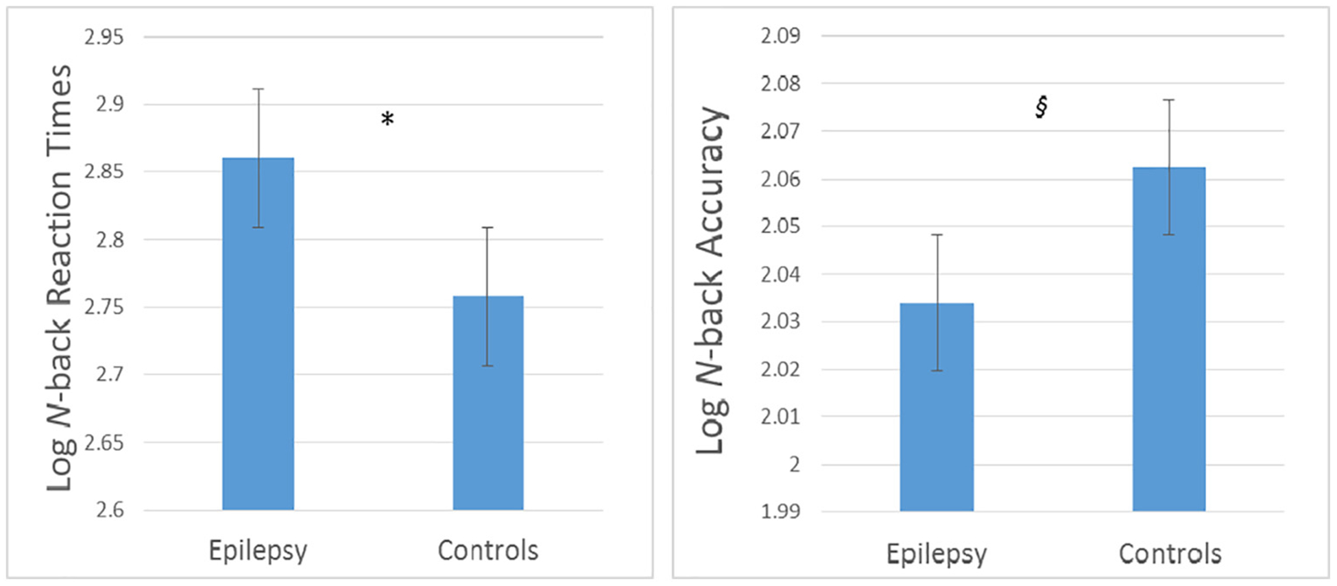
Experimental design: fMRI was conducted on 15 children with TLE and 15 healthy controls (age: 8-16 years) while performing the N-back task in order to assess deactivation of the DMN. N-back accuracy, N-back reaction time, and neuropsychological tests of executive function (Delis-Kaplan Executive Function System [D-KEFS] Color-Word Interference and Card Sort tests) were also assessed.
Principal observations: During the N-back task, children with TLE exhibited significantly less deactivation of the DMN, primarily in the precuneus/posterior cingulate cortex compared with controls. These alterations significantly correlated with N-back behavioral findings and D-KEFS results.
Conclusions: Children with TLE exhibit executive dysfunction which correlates with DMN alterations. These findings suggest that the level of deactivation of specific functional networks may contribute to cognitive impairment in children with TLE. The findings also indicate that children with TLE have network alterations in extratemporal lobe brain regions.
Keywords: Default mode network; Executive dysfunction; N-back task; Neuropsychological testing; Pediatric; Temporal lobe epilepsy.
Copyright © 2019 Elsevier Inc. All rights reserved.
Conflict of interest statement
Conflicts of interest
We have no conflicts of interest to report.
Figures


References
-
- Sala-Llonch R, Pena-Gomez C, Arenaza-Urquijo EM, Vidal-Piñeiro D, Bargalló N, Junqué C, et al. Brain connectivity during resting state and subsequent working memory task predicts behavioural performance. Cortex 2012;48:1187–96. - PubMed
-
- Buckner RL, Andrews-Hanna JR, Schacter DL. The brain’s default network: anatomy, function, and relevance to disease. Ann N Y Acad Sci 2008;1124:1–38. - PubMed
-
- Gauffin H, van Ettinger-Veenstra H, Landtblom A, Ulrici D, McAllister A, Karlsson T, et al. Impaired language function in generalized epilepsy: inadequate suppression of the default mode network. Epilepsy Behav 2013;28:26–35. - PubMed
