Review
. 2019 Mar 22;20(6):1456.
doi: 10.3390/ijms20061456.

Human Vascular Pericytes and Cytomegalovirus Pathobiology

Affiliations
Review

Human Vascular Pericytes and Cytomegalovirus Pathobiology

Donald J Alcendor. Int J Mol Sci. .

Abstract

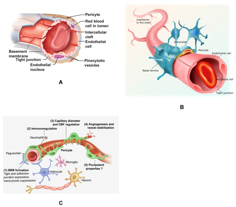
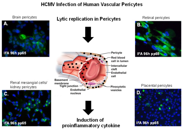
Pericytes are multipotent cells of the vascular system with cytoplasmic extensions proximal to endothelial cells that occur along the abluminal surface of the endothelium. The interactions between endothelial cells and pericytes are essential for proper microvascular formation, development, stabilization, and maintenance. Pericytes are essential for the regulation of paracellular flow between cells, transendothelial fluid transport, angiogenesis, and vascular immunosurveillance. They also influence the chemical composition of the surrounding microenvironment to protect endothelial cells from potential harm. Dysregulation or loss of pericyte function can result in microvascular instability and pathological consequences. Human pericytes have been shown to be targets for human cytomegalovirus (HCMV) infection and lytic replication that likely contribute to vascular inflammation. This review focuses on human vascular pericytes and their permissiveness for HCMV infection. It also discusses their implication in pathogenesis in the blood⁻brain barrier (BBB), the inner blood⁻retinal barrier (IBRB), the placenta⁻blood barrier, and the renal glomerulus as well as their potential role in subclinical vascular disease.

Keywords: HCMV; brain; cytomegalovirus; endothelial; inflammation; ocular; pericyte; placenta; renal; vascular.


Conflict of interest statement

The author declares no conflict of interest.

Figures

Figure 1
Figure 1
Brain capillaries featuring pericytes. (A) Cross-section of a brain capillary illustrating the capillary basement membrane, as well as the endothelial cells and brain pericytes abluminal to endothelial cells sharing a contiguous basement membrane. (B). The neurovascular unit (NVU) of the blood–brain barrier (BBB) featuring capillary endothelial cells with tight junction, pericytes and astrocytes. (C) The NVU and other brain parenchymal cells, including neurons and microglia. Important functions of pericytes are also shown listed 1–5.
Figure 2
Figure 2
Pericyte morphology and cultivation. (A) Transmission electron micrograph of the abluminal surface of a rat brain capillary showing the arrangement of pericytes with long cytoplasmic extensions. (B) Cross-section of a rat brain capillary showing microvascular endothelial cells and pericytes. (C) Subconfluent culture of normal primary human brain pericytes with long cytoplasmic extensions. (D) Confluent culture of normal primary human brain pericytes. Images (A) and (B) (modified with permission from Pearson Education Inc. (unpublished data). Phase images for (C) and (D) were taken on a Nikon TE2000S microscope mounted with a charge-coupled device (CCD) camera at ×200 magnification.
Figure 3
Figure 3
Vascular pericytes and HCMV infectivity. HCMV lytic infection of human pericytes. Black arrows point to a cross-section of a capillary showing HCMV dissemination in vasculature with virus infection concentrated in pericytes leading to the induction proinflammatory cytokines. (A) Immunofluorescent staining of primary human brain pericytes in with SBCMV 96 h after infection using a mouse monoclonal antibody to the HCMV pp65 tegument protein. (B) Immunostaining of primary human retinal pericytes infected with SBCMV at 96 h stained with the HCMV pp65 antibody. (C) Immunostaining of primary human renal mesangial cells infected with HCMV at 96 h and stained with the HCMV pp65 antibody. (D) Immunostaining of primary human placental pericytes infected with SBCMV at 96 h stained with the HCMV pp65 antibody. All images were taken on a Nikon TE2000S microscope mounted with a CCD camera at ×200 magnification. For fluorescent images, 4′,6-diamidino-2-phenylindole (DAPI) was used to stain the nuclei (blue).

References

    1. Eberth C.J. Handbuch der Lehre von der Gewegen des Menschen und der Tiere. Volume 1 Engelmann; Leipzig, Germany: 1871.
    1. Rouget C. Me’moire sur le de´veloppement, la structure et les propriete´s physiologiques des capillaires sanguins et lymphatiques. Arch. Physiol. Norm Path. 1873;5:603–663.
    1. Zimmermann K.W. Der feinere Bau der Blutkapillaren. Z. Anat. Entwicklungsgesch. 1923;68:29–109. doi: 10.1007/BF02593544. - DOI
    1. Attwell D., Mishra A., Hall C.N., O’Farrell F.M., Dalkara T. What is a pericyte? J. Cereb. Blood Flow Metab. 2016;2:451–455. doi: 10.1177/0271678X15610340. - DOI - PMC - PubMed
    1. Sims D.E. The pericyte—A review. Tissue Cell. 1986;18:153–174. doi: 10.1016/0040-8166(86)90026-1. - DOI - PubMed

MeSH terms