. 2019 Mar 22;20(6):1458.
doi: 10.3390/ijms20061458.

Change in Renal Glomerular Collagens and Glomerular Filtration Barrier-Related Proteins in a Dextran Sulfate Sodium-Induced Colitis Mouse Model

Affiliations

Change in Renal Glomerular Collagens and Glomerular Filtration Barrier-Related Proteins in a Dextran Sulfate Sodium-Induced Colitis Mouse Model

Chia-Jung Chang et al. Int J Mol Sci. .

Abstract

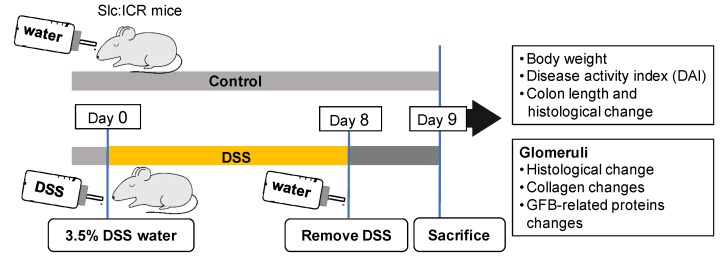
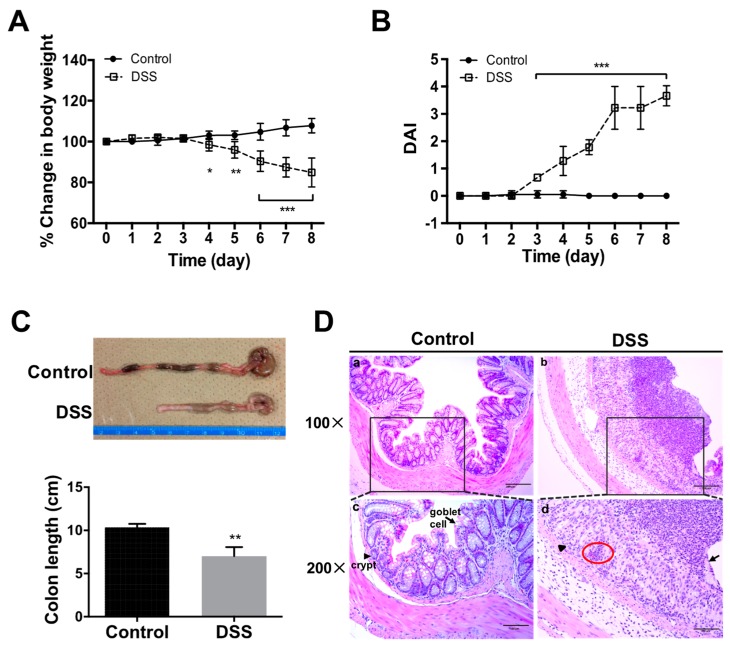
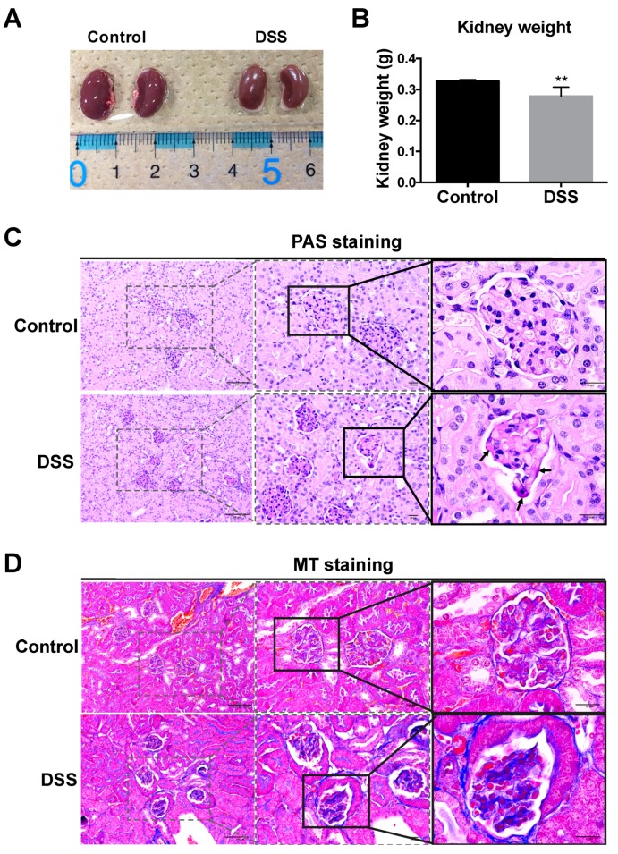
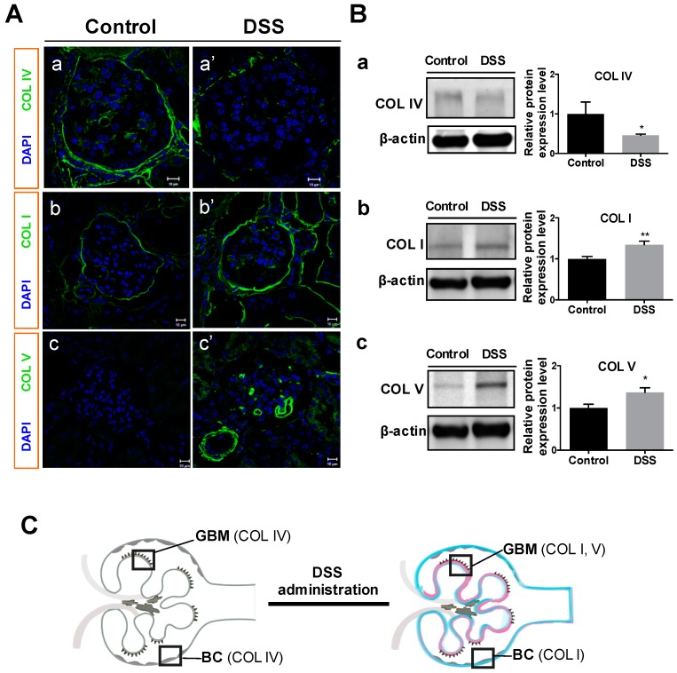
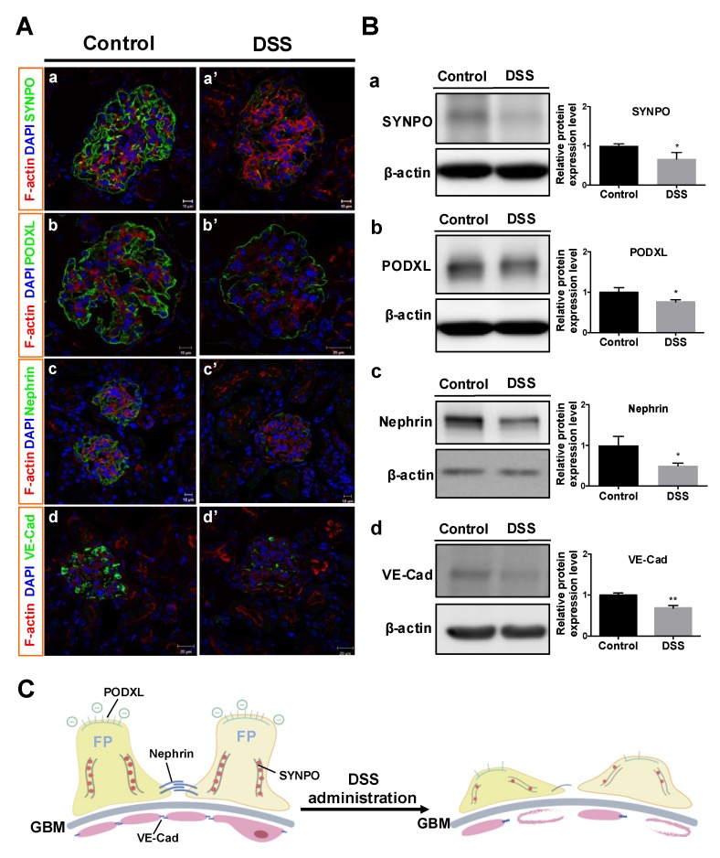
Renal disease is not rare among patients with inflammatory bowel disease (IBD) and is gaining interest as a target of research. However, related changes in glomerular structural have rarely been investigated. This study was aimed at clarifying the changes in collagens and glomerular filtration barrier (GFB)-related proteins of glomeruli in a dextran sulfate sodium (DSS)-induced colitis mouse model. Acute colitis was induced by administering 3.5% DSS in Slc:ICR strain mice for eight days. Histological changes to glomeruli were examined by periodic acid-Schiff (PAS) and Masson's trichrome staining. Expressions of glomerular collagens and GFB-related proteins were analyzed by immunofluorescent staining and Western blot analysis. DSS-colitis mice showed an elevated disease activity index (DAI), colon shortening, massive cellular infiltration and colon damage, confirming that DSS-colitis mice can be used as an IBD animal model. DSS-colitis mice showed increased glycoprotein and collagen deposition in glomeruli. Interestingly, we observed significant changes in glomerular collagens, including a decrease in type IV collagen, and an increment in type I and type V collagens. Moreover, declined GFB-related proteins expressions were detected, including synaptopodin, podocalyxin, nephrin and VE-cadherin. These results suggest that renal disease in DSS-colitis mice might be associated with changes in glomerular collagens and GFB-related proteins. These findings are important for further elucidation of the clinical pathological mechanisms underlying IBD-associated renal disease.

Keywords: DSS-colitis; glomerular filtration barrier (GFB); inflammatory bowel disease (IBD); type I collagen; type IV collagen; type V collagen.


Conflict of interest statement

The authors declare that they have no conflicts of interest, financial or otherwise, regarding this article.

Figures

Figure 1
Figure 1
Investigating mouse glomerular structural changes associated with dextran sulfate sodium (DSS)-induced colitis. Slc:ICR mice were administered 3.5% DSS in drinking water for eight days, then allowed intake of filtered water on Day 8. Control mice were given filtered water. All mice were sacrificed on Day 9 and further assessments were performed. Abbreviation: GFB, glomerular filtration barrier.
Figure 2
Figure 2
Macro- and microscopic changes to bowel in mice with DSS-induced colitis. Changes in body weight (A) and disease activity index (DAI) (B) were evaluated daily. Colon length was measured after sacrifice (C). Hematoxylin and eosin (HE) staining (D) showed distortion of crypts (arrowhead), loss of goblet cells (arrow), and infiltration of inflammatory cells (red circle) in colon sections from DSS-treated mice. All values are given as mean ± SEM (n = 6 mice); * p < 0.05, ** p < 0.01, and *** p < 0.001 vs. control. Scale bars: 200 μm (a,b); and 100 μm (c,d).
Figure 3
Figure 3
Macro- and microscopic changes to the kidney and glomeruli in mice after DSS administration. Mouse kidney appearance (A) and weight (B) were determined at harvest. Histological manifestations were determined by staining with periodic acid-Schiff (PAS) to assess the basement membrane of glomeruli (C), and Masson’s trichrome (MT) staining to assess collagen deposition (D), respectively. Compared to control mice, glomerular accumulation of PAS-positive matrix (arrow) was prominent in DSS-treated mice (C). Blue staining indicates the presence of collagen fibers in tissues (D). All values are given as mean ± SEM (n = 6 mice); ** p < 0.01 vs. control. Scale bars: 100 μm and 20 μm.
Figure 4
Figure 4
Changes in glomerular collagens in mice after DSS administration. Immunofluorescent microscopy (A) and Western blot analysis of protein expression (B) for type IV collagen (COL IV; A-a, A-a’; B-a; 160–190 kDa), type I collagen (COL I; A-b, A-b’; B-b; 150 kDa), and type V collagen (COL V; A-c, A-c’; B-c; 220 kDa) were conducted for control and DSS-colitis mice. Representative bands (B, left) and relative band intensity ratios were analyzed (B, right). (C) Illustration of glomerular collagens changes in this study. All values are means ± SEM (n = 6); * p < 0.05 and ** p < 0.01 vs. control. Scale bars = 10 μm. Abbreviations: GBM, glomerular basement membrane; BC, Bowman’s capsule.
Figure 5
Figure 5
Changes in GFB-related proteins in mice after DSS administration. Immunofluorescent microscopy (A) and Western blot analysis of protein expression (B) against synaptopodin (A-a, A-a’; B-a; 100 kDa), podocalyxin (A-b, A-b’; B-b; 130 kDa), nephrin (A-c, A-c’; B-c; 185 kDa) and VE-cadherin (A-d, A-d’; B-d; 130 kDa) in glomeruli were conducted for control and DSS-colitis mice. (B) Representative bands (left), and relative band intensity ratios (right) were analyzed. (C) Illustration of GFB-related proteins changes in this study. All values are means ± SEM (n = 6), * p < 0.05 and ** p < 0.01 vs. control. Scale bars = 10 μm (A-a, A-a’, A-b, A-c, A-c’); 20 μm (A-b’, A-d, A-d’). Abbreviation: SYNPO, synaptopodin; PODXL, podocalyxin; VE-Cad, VE-cadherin; FP, foot processes.

References

    1. Sartor R.B. Current concepts of the etiology and pathogenesis of ulcerative colitis and Crohn’s disease. Gastroenterol. Clin. N. Am. 1995;24:475–507. - PubMed
    1. Ricart E., Panaccione R., Loftus E.V., Tremaine W.J., Harmsen W.S., Zinsmeister A.R., Sandborn W.J. Autoimmune disorders and Extraintestinal manifestations in First-degree familial and sporadic inflammatory bowel disease. Inflamm. Bowel Dis. 2004;10:207–214. doi: 10.1097/00054725-200405000-00005. - DOI - PubMed
    1. Christodoulou D.K., Katsanos K.H., Kitsanou M., Stergiopoulou C., Hatzis J., Tsianos E.V. Frequency of extraintestinal manifestations in patients with inflammatory bowel disease in northwest Greece and review of the literature. Dig. Liver Dis. 2002;34:781–786. doi: 10.1016/S1590-8658(02)80071-8. - DOI - PubMed
    1. Ambruzs J.M., Walker P.D., Larsen C.P. The histopathologic spectrum of kidney biopsies in patients with inflammatory bowel disease. Clin. J. Am. Soc. Nephrol. 2014;9:265–270. doi: 10.2215/CJN.04660513. - DOI - PMC - PubMed
    1. Ambruzs J.M., Larsen C.P. Renal Manifestations of Inflammatory Bowel Disease. Rheum. Dis. Clin. N. Am. 2018;44:699–714. doi: 10.1016/j.rdc.2018.06.007. - DOI - PubMed

LinkOut - more resources