. 2019 Mar 23;20(6):1473.
doi: 10.3390/ijms20061473.

Intravenous Immunoglobulin Therapy Eliminates Candida albicans and Maintains Intestinal Homeostasis in a Murine Model of Dextran Sulfate Sodium-Induced Colitis

Affiliations

Intravenous Immunoglobulin Therapy Eliminates Candida albicans and Maintains Intestinal Homeostasis in a Murine Model of Dextran Sulfate Sodium-Induced Colitis

Rogatien Charlet et al. Int J Mol Sci. .

Abstract

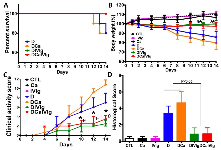
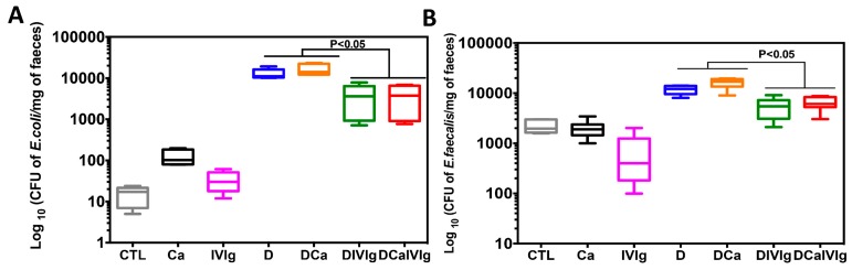
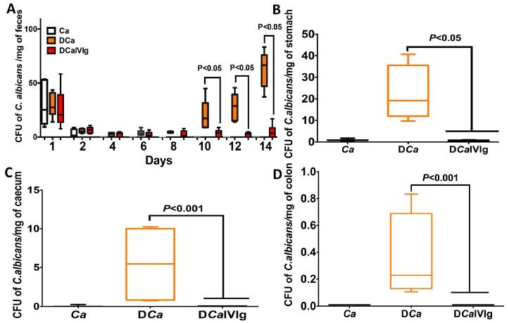
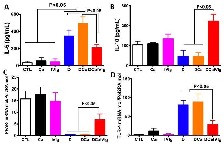
Intravenous immunoglobulin (IVIg) therapy has diverse anti-inflammatory and immunomodulatory effects and has been employed successfully in autoimmune and inflammatory diseases. The role of IVIg therapy in the modulation of intestinal inflammation and fungal elimination has not been yet investigated. We studied IVIg therapy in a murine model of dextran sulfate sodium (DSS)-induced colitis. Mice received a single oral inoculum of Candida albicans and were exposed to DSS treatment for 2 weeks to induce colitis. All mice received daily IVIg therapy starting on day 1 for 7 days. IVIg therapy not only prevented a loss of body weight caused by the development of colitis but also reduced the severity of intestinal inflammation, as determined by clinical and histological scores. IVIg treatment significantly reduced the Escherichia coli, Enterococcus faecalis, and C. albicans populations in mice. The beneficial effects of IVIg were associated with the suppression of inflammatory cytokine interleukin (IL)-6 and enhancement of IL-10 in the gut. IVIg therapy also led to an increased expression of peroxisome proliferator-activated receptor gamma (PPARγ), while toll-like receptor 4 (TLR-4) expression was reduced. IVIg treatment reduces intestinal inflammation in mice and eliminates C. albicans overgrowth from the gut in association with down-regulation of pro-inflammatory mediators combined with up-regulation of anti-inflammatory cytokines.

Keywords: Candida albicans; Enterococcus faecalis; Escherichia coli; colitis; cytokines; dextran sulfate sodium; inflammation; intravenous immunoglobulin G; mice.


Conflict of interest statement

The authors declare no conflict of interest.

Figures

Figure 1
Figure 1
Effect of intravenous immunoglobulin (IVIg) treatment on the survival of mice, body weight, and inflammatory scores in the dextran sulfate sodium (DSS)-induced colitis model. (A) Mouse survival. Results are expressed as percent survival from the time of C. albicans challenge and DSS treatment. CTL, Ca, IVIg or D correspond to control groups receiving water, C. albicans, IVIg treatment or DSS, respectively. DCa corresponds to mice receiving C. albicans and treated with DSS. DIVIg corresponds to mice receiving DSS and treated with IVIg. DCaIVIg represents mice receiving C. albicans challenged with IVIg and treated with DSS. No mouse mortality was recorded in the Ca or IVIg groups, while DSS treatment induced 20% mouse mortality in groups D and DCa; (B) Body weight. Results are expressed as a percent; (C) Clinical analysis of DSS-induced colitis in mice after IVIg treatment. Clinical score was determined by assessing weight loss, change in stool consistency, and presence of gross bleeding. The clinical score ranged from 0 to 12 (each value corresponds to the mean value over 14 days per group). * p < 0.05 for DCaIVIg mice vs. DCa and D mice; (D) Histological scores. Scores range from 0 (no changes) to six (extensive cell infiltration and tissue damage). Data are the mean ± SD of 14 mice per group.
Figure 2
Figure 2
Histological analysis of colons from mice with dextran sulfate sodium (DSS)-induced colitis. Panels (a,b) display sections from mice that received C. albicans; panels (c,d) correspond to C. albicans + DSS; and panels (e,f) denote colon sections from mice that received C. albicans + DSS + intravenous immunoglobulin (IVIg). C. albicans alone did not induce any epithelial injury or an inflammatory cell infiltrate in the colon wall structure. Colon sections from DCa mice show an inflammatory cell infiltrate in colon wall structures and massive tissue destruction (asterisk), while colon sections from DCaIVIg mice show minimal tissue damage. The scale bars represent 50 µm (a,c,e) and 10 µm (b,d,f).
Figure 3
Figure 3
Determination of viable fecal aerobic bacteria in mice with colitis and treated with intravenous immunoglobulin (IVIg) (A,B) The number of E. coli and E. faecalis colonies recovered from colonic luminal contents. Data are expressed as box-and-whiskers plots, with min to the max range as whiskers. CFU: colony forming units.
Figure 4
Figure 4
Evaluation of C. albicans colonization in various organs following intravenous immunoglobulin (IVIg) treatment of mice with dextran sulfate sodium (DSS)-induced colitis. (A) The number of C. albicans colony forming units (CFU) recovered from stools. Data are expressed as box-and-whiskers plots, with min to the max range as whiskers. (BD) The number of C. albicans CFU recovered from the stomach, cecum, and colon. Data are expressed as box-and-whiskers plots, with min to the max range as whiskers.
Figure 5
Figure 5
Cytokine and receptor expression after intravenous immunoglobulin (IVIg) treatment of mice with C. albicans and dextran sulfate sodium (DSS)-induced colitis. (A,B) Protein levels of interleukin (IL)-6 and IL-10 in mouse colons. (C,D) Relative expression of peroxisome proliferator-activated receptor gamma (PPARγ) and toll-like receptor 4 (TLR-4) mRNA in mouse colons. Data are the mean ± SD of 14 mice per group.

Similar articles

Cited by

References

    1. Poulain D. Candida albicans, plasticity and pathogenesis. Crit. Rev. Microbiol. 2015;41:208–217. doi: 10.3109/1040841X.2013.813904. - DOI - PubMed
    1. Kullberg B.J., Arendrup M.C. Invasive candidiasis. N. Engl. J. Med. 2015;373:1445–1456. doi: 10.1056/NEJMra1315399. - DOI - PubMed
    1. Gow N.A., van de Veerdonk F.L., Brown A.J., Netea M.G. Candida albicans morphogenesis and host defence: Discriminating invasion from colonization. Nat. Rev. Microbiol. 2012;10:112–122. doi: 10.1038/nrmicro2711. - DOI - PMC - PubMed
    1. Sendid B., Dotan N., Nseir S., Savaux C., Vandewalle P., Standaert A., Zerimech F., Guery B.P., Dukler A., Colombel J.F., et al. Antibodies against glucan, chitin, and saccharomyces cerevisiae mannan as new biomarkers of candida albicans infection that complement tests based on c. Albicans mannan. Clin. Vaccine Immunol. 2008;15:1868–1877. doi: 10.1128/CVI.00200-08. - DOI - PMC - PubMed
    1. Frank D.N., St Amand A.L., Feldman R.A., Boedeker E.C., Harpaz N., Pace N.R. Molecular-phylogenetic characterization of microbial community imbalances in human inflammatory bowel diseases. Proc. Natl. Acad. Sci. USA. 2007;104:13780–13785. doi: 10.1073/pnas.0706625104. - DOI - PMC - PubMed

MeSH terms

LinkOut - more resources