Review
. 2018 Jan-Dec:10:1759091418818261.
doi: 10.1177/1759091418818261.

Mitochondrial Substrate-Level Phosphorylation as Energy Source for Glioblastoma: Review and Hypothesis

Affiliations
Review

Mitochondrial Substrate-Level Phosphorylation as Energy Source for Glioblastoma: Review and Hypothesis

Christos Chinopoulos et al. ASN Neuro. 2018 Jan-Dec.

Abstract

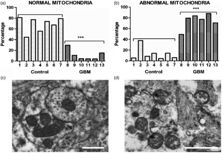
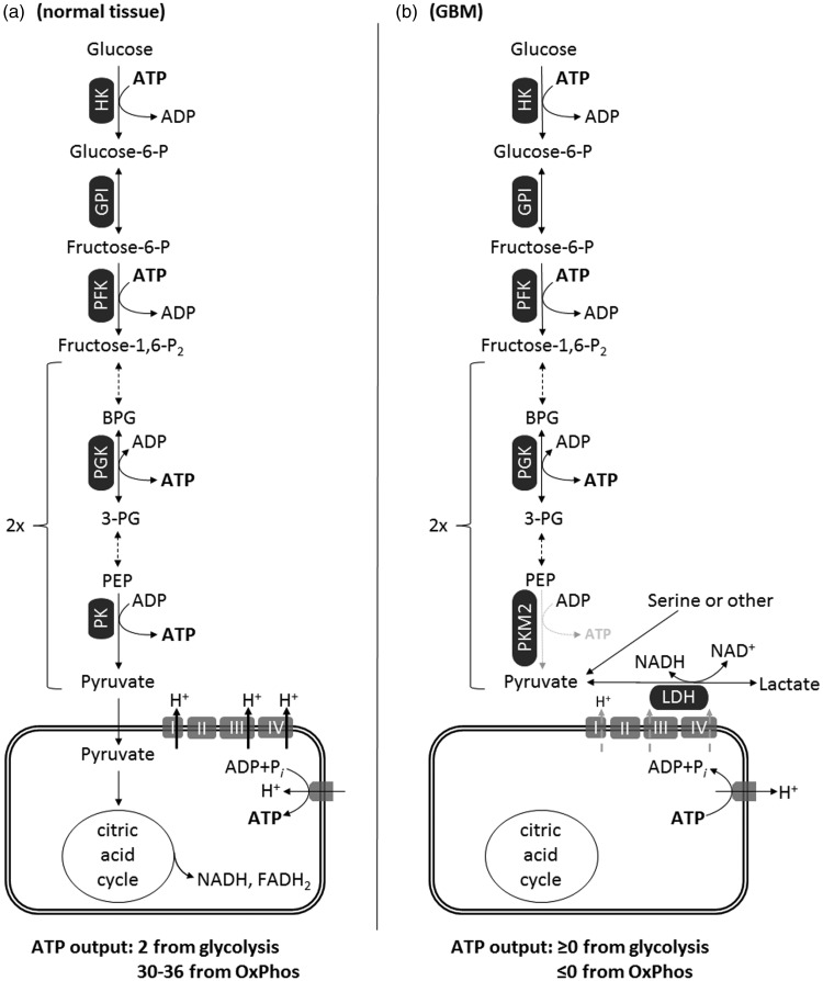
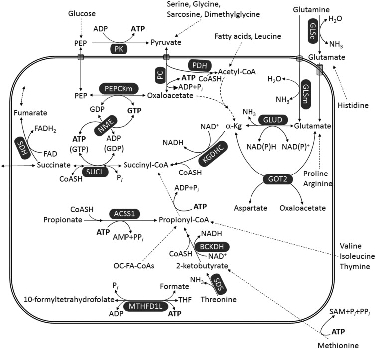
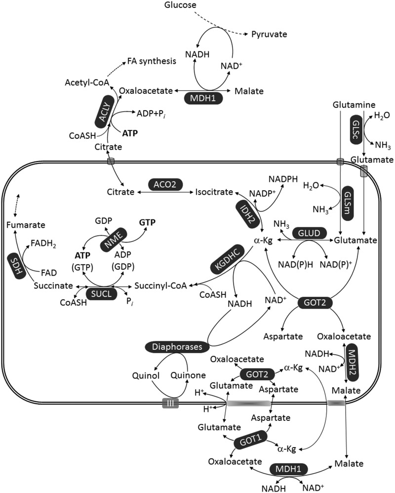
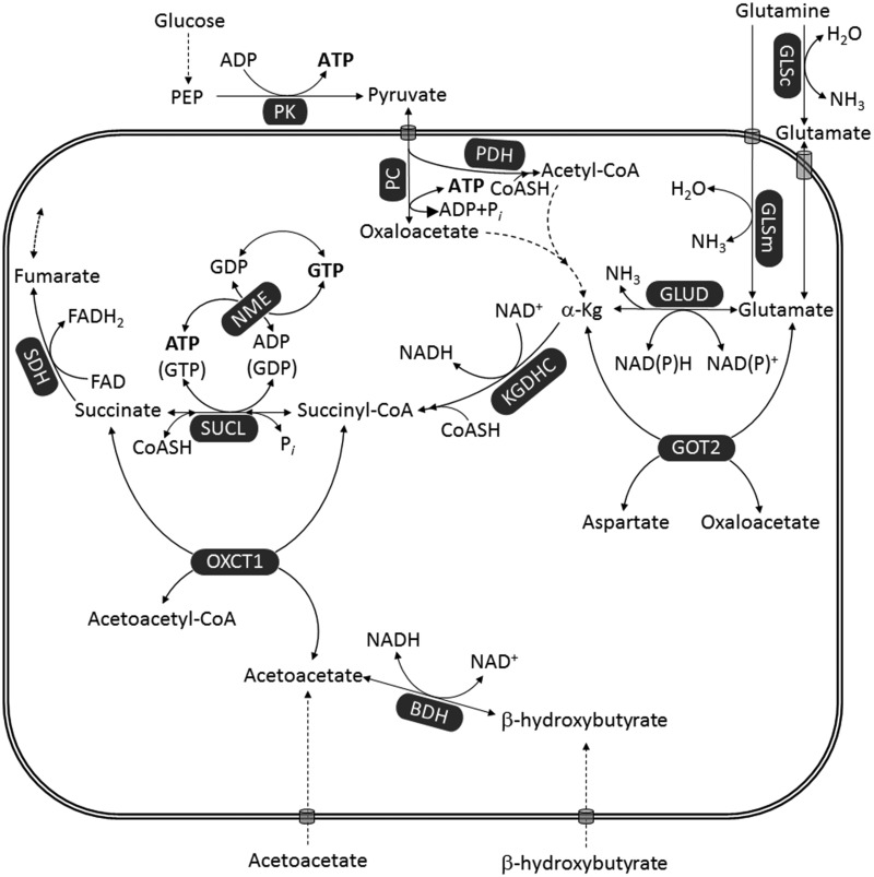
Glioblastoma multiforme (GBM) is the most common and malignant of the primary adult brain cancers. Ultrastructural and biochemical evidence shows that GBM cells exhibit mitochondrial abnormalities incompatible with energy production through oxidative phosphorylation (OxPhos). Under such conditions, the mitochondrial F0-F1 ATP synthase operates in reverse at the expense of ATP hydrolysis to maintain a moderate membrane potential. Moreover, expression of the dimeric M2 isoform of pyruvate kinase in GBM results in diminished ATP output, precluding a significant ATP production from glycolysis. If ATP synthesis through both glycolysis and OxPhos was impeded, then where would GBM cells obtain high-energy phosphates for growth and invasion? Literature is reviewed suggesting that the succinate-CoA ligase reaction in the tricarboxylic acid cycle can substantiate sufficient ATP through mitochondrial substrate-level phosphorylation (mSLP) to maintain GBM growth when OxPhos is impaired. Production of high-energy phosphates would be supported by glutaminolysis-a hallmark of GBM metabolism-through the sequential conversion of glutamine → glutamate → alpha-ketoglutarate → succinyl CoA → succinate. Equally important, provision of ATP through mSLP would maintain the adenine nucleotide translocase in forward mode, thus preventing the reverse-operating F0-F1 ATP synthase from depleting cytosolic ATP reserves. Because glucose and glutamine are the primary fuels driving the rapid growth of GBM and most tumors for that matter, simultaneous restriction of these two substrates or inhibition of mSLP should diminish cancer viability, growth, and invasion.

Keywords: Warburg; bioenergetics; gliomas; therapies.


Figures

Figure 1.
Figure 1.
Morphological abnormalities in GBM mitochondria; (a) to (d) (distributed under a Creative Commons license) were reproduced from Deighton et al. (2014). The morphology of 150 mitochondria was assessed in six GBM samples and in seven peritumoural control samples using electron microscopy. (a) Percentage of normal mitochondria where cristae were visible throughout the mitochondria in peritumoural control and GBM samples (each bar represents one sample; ***p value = .0001); (b) Percentage of abnormal mitochondria where cristae were sparse and abnormal in peritumoural control and GBM samples (***p value = .0001). (c and d) Representative electron microscopy images of normal and abnormal mitochondria, respectively. The scale bars represent 0.5 µm. Cristolysis was significantly greater in mitochondria from GBM than from control brain. The possibility cannot be excluded that the abnormal GBM mitochondria shown in panel (d) originate from areas close to necrotic palisades and might not be representative of mitochondria in all types GBM cells. GBM = glioblastoma multiforme.
Figure 2.
Figure 2.
Energetics of glycolysis and OxPhos in normal tissue (a) versus GBM (b). In GBM, the diminished activity of dimeric PKM2 to generate ATP yields a net ≥0 ATP output from glycolysis. Also, because GBM mitochondria exhibit a dysfunctional ETC and a reverse-operating F0-F1 ATP synthase, they are ATP consumers (≤0 ATP output). OxPhos in GBM mitochondria may operate at a diminished rate, and this is depicted by the gray-dashed arrows traversing complexes I, III, and IV implying diminished ability for proton expulsion. FADH = reduced form of flavin adenine dinucleotide; GBM = glioblastoma multiforme; GPI = phospohexose isomerase; HK = hexokinase (any isoform); LDH = lactate dehydrogenase; PEP = phosphoenolpyruvate; PFK = phosphofructokinase; PGK = phosphoglycerate kinase; PK = pyruvate kinase; PKM2 = pyruvate kinase M2 isoform, dimeric; BPG = biphosphoglycerate; 3-PG = 3-phosphoglycerate.
Figure 3.
Figure 3.
Pathways leading to ATP (or GTP) generation at the substrate level. Dashed arrows imply multiple enzymatic steps (omitted for clarity). Reduction of NAD+ to NADH by PDH is omitted for uncluttering the figure. α-Kg = α-ketoglutarate; ACSS1 = acetyl-coenzyme A synthetase 2-like, mitochondrial; BCKDH = branched-chain α-ketoacid dehydrogenase; GLSc = glutaminase, cytosolic; GLSm = glutaminase, mitochondrial; GLUD = glutamate dehydrogenase; GOT2 = aspartate aminotransferase; KGDHC = α-ketoglutarate dehydrogenase complex; MTHFD1L = monofunctional C1-tetrahydrofolate synthase; NME = nucleoside diphosphate kinase; OC-FA = odd-chain fatty acid; Pi = inorganic phosphate; PPi = pyrophosphate; PC = pyruvate carboxylase; PDH = pyruvate dehydrogenase; PEP = phosphoenolpyruvate; PEPCKm = mitochondrial phosphoenolpyruvate carboxykinase; PK = pyruvate kinase; SAM = S-adenosylmethionine; SDH = succinate dehydrogenase; SDS = serine dehydratase; SUCL = succinate-CoA ligase; THF = tetrahydrofolate.
Figure 4.
Figure 4.
Provision of NAD+ in the matrix of mitochondria in the absence of OxPhos. Enzyme annotations as in Figures 2 and 3. α-Kg = α-ketoglutarate; ACO2 = aconitase; ACLY = ATP citrate lyase; FA = fatty acid; FAD = flavin adenine dinucleotide; GLSc = glutaminase, cytosolic; GLSm = glutaminase, mitochondrial; GLUD = glutamate dehydrogenase; GOT2 = aspartate aminotransferase; IDH2 = NADP+-dependent isocitrate dehydrogenase; KGDHC = α-ketoglutarate dehydrogenase complex; MDH = malate dehydrogenase; NME = nucleoside diphosphate kinase; SDH = succinate dehydrogenase; SUCL = succinate-CoA ligase.
Figure 5.
Figure 5.
Ketone bodies bypass mSLP. Enzyme annotations as for Figures 2, 3, and 4. BDH = β-hydroxybutyrate dehydrogenase; FAD = flavin adenine dinucleotide; GLSc = glutaminase, cytosolic; GLSm = glutaminase, mitochondrial; GLUD = glutamate dehydrogenase; GOT2 = aspartate aminotransferase; KGDHC = α-ketoglutarate dehydrogenase complex; NME = nucleoside diphosphate kinase; OXCT1 = succinyl-CoA:3-ketoacid coenzyme A transferase 1; PC = pyruvate carboxylase; PDH = pyruvate dehydrogenase; PEP = phosphoenolpyruvate; PK = pyruvate kinase; SUCL = succinate-CoA ligase.

References

    1. Adam J., et al. (2011). Renal cyst formation in Fh1-deficient mice is independent of the Hif/Phd pathway: Roles for fumarate in KEAP1 succination and Nrf2 signaling. Cancer Cell, 20, 524–537. - PMC - PubMed
    1. Ahluwalia G. S., Grem J. L., Hao Z., Cooney D. A. (1990). Metabolism and action of amino acid analog anti-cancer agents. Pharmacol Ther, 46, 243–271. - PubMed
    1. Ahmadloo N., Kani A. A., Mohammadianpanah M., Nasrolahi H., Omidvari S., Mosalaei A., Ansari M. (2013). Treatment outcome and prognostic factors of adult glioblastoma multiforme. J Egypt Natl Canc Inst, 25, 21–30. - PubMed
    1. Aiken K. J., Bickford J. S., Kilberg M. S., Nick H. S. (2008). Metabolic regulation of manganese superoxide dismutase expression via essential amino acid deprivation. J Biol Chem, 283, 10252–10263. - PMC - PubMed
    1. Alam N. A., et al. (2003). Genetic and functional analyses of FH mutations in multiple cutaneous and uterine leiomyomatosis, hereditary leiomyomatosis and renal cancer, and fumarate hydratase deficiency. Hum Mol Genet, 12, 1241–1252. - PubMed

Publication types