. 2019 May;25(5):276-287.
doi: 10.1089/ten.TEC.2018.0339.

Fabrication and Characterization of Electrospun Decellularized Muscle-Derived Scaffolds

Affiliations

Fabrication and Characterization of Electrospun Decellularized Muscle-Derived Scaffolds

Mollie M Smoak et al. Tissue Eng Part C Methods. 2019 May.

Abstract

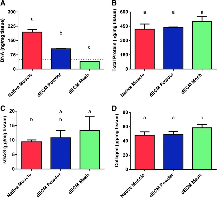
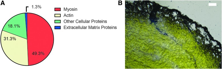
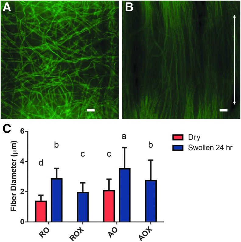
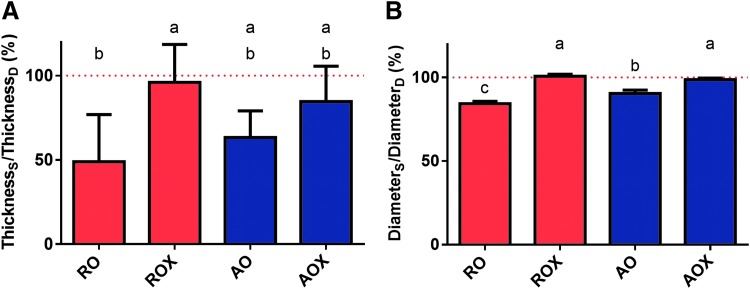
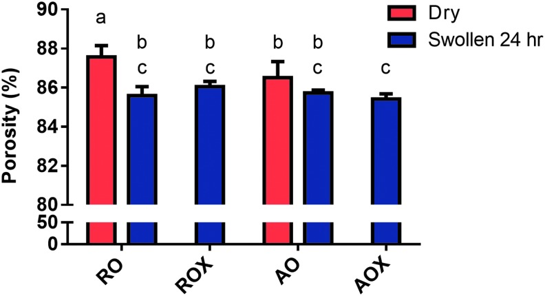
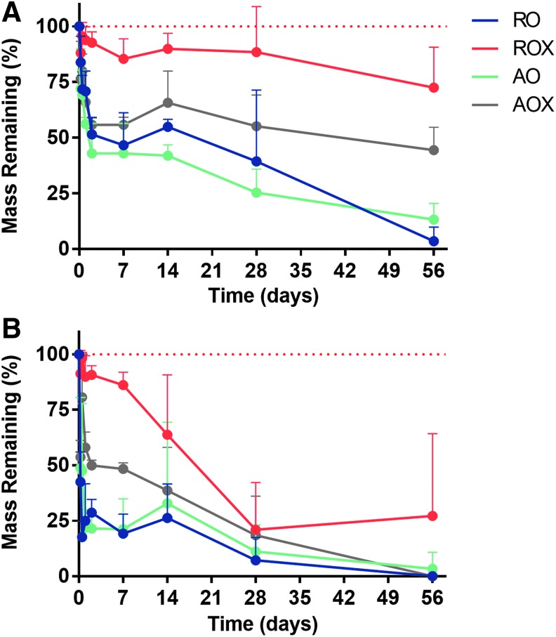
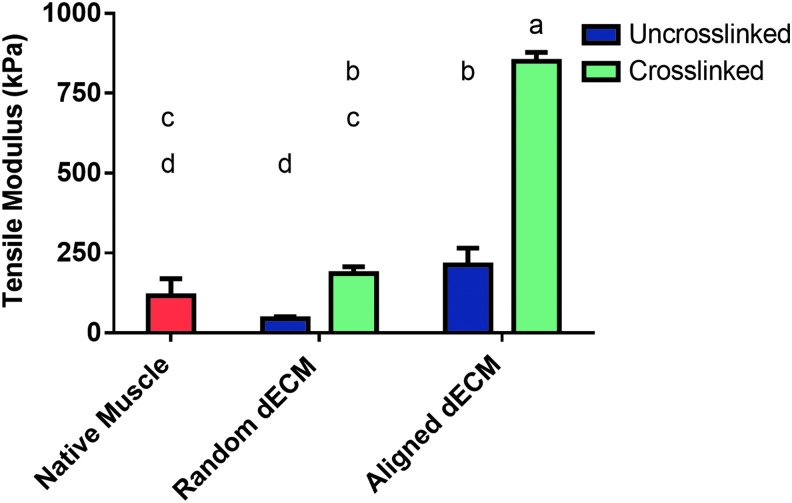
Although skeletal muscle has a high potential for self-repair, volumetric muscle loss can result in impairment beyond the endogenous regenerative capacity. There is a clinical need to improve on current clinical treatments that fail to fully restore the structure and function of lost muscle. Decellularized extracellular matrix (dECM) scaffolds have been an attractive platform for regenerating skeletal muscle, as dECM contains many biochemical cues that aid in cell adhesion, proliferation, and differentiation. However, there is limited capacity to tune physicochemical properties in current dECM technologies to improve outcome. In this study, we aim to create a novel, high-throughput technique to fabricate dECM scaffolds with tunable physicochemical properties while retaining proregenerative matrix components. We demonstrate a successful decellularization protocol that effectively removes DNA. We also identified key steps for the successful production of electrospun muscle dECM without the use of a carrier polymer; electrospinning allows for rapid scaffold fabrication with high control over material properties, which can be optimized to mimic native muscle. To this end, fiber orientation and degree of crosslinking of these dECM scaffolds were modulated and the corollary effects on fiber swelling, mechanical properties, and degradation kinetics were investigated. Beyond application in skeletal muscle, the versatility of this technology has the potential to serve as a foundation for dECM scaffold fabrication in a variety of tissue engineering applications.

Keywords: decellularized muscle; electrospinning; extracellular matrix; skeletal muscle.


Conflict of interest statement

No competing financial interests exist.

Figures

<b>FIG. 1.</b>
FIG. 1.
Schematic of decellularization and production of dECM electrospun fibers. (A) Skeletal muscle was harvested from the hind limbs of New Zealand White rabbits and removed of all visible fascia and connective tissue. (B) Skeletal muscle was exposed to a number of reagents to remove cellular material from the extracellular matrix. (C) After homogenization and drying, dECM was electrospun without the use of a carrier polymer to form dECM fibers. The scale bar represents 10 μm. dECM, decellularized extracellular matrix.
<b>FIG. 2.</b>
FIG. 2.
Biochemical analysis of untreated (native) skeletal muscle, dECM powder, and dECM electrospun mesh. (A) DNA was quantified via PicoGreen assay. A dashed red line represents 50 ng/mg of tissue, which is the current recommended upper limit for decellularized material. (B) Total protein was quantified via BCA assay. (C) sGAG content was quantified via DMMB assay, and (D) collagen content was quantified via hydroxyproline assay. Shared letters indicate no significant difference between groups (n = 3, p < 0.05). BCA, bicinchoninic acid; sGAG, sulfated glycosaminoglycans; DMMB, 1,9-dimethylmethylene blue.
<b>FIG. 3.</b>
FIG. 3.
Identification of proteins in decellularized muscle. Protein identification was performed on electrospun dECM scaffolds using LC-MS/MS (A). Decellularized muscle was also evaluated histologically for collagen using an Alcian blue stain (B).
<b>FIG. 4.</b>
FIG. 4.
Analysis of fiber swelling. (A) Representative images of swollen random (RO) dECM fibers and (B) swollen aligned (AO) dECM fibers were taken. The scale bar represents 10 μm. The arrow represents the direction of fiber orientation. (C) Fiber diameter was measured dry (red bars) and after 24 h of swelling in PBS (blue bars). Shared letters indicate no significant difference between groups (n ≥ 50, p < 0.05). PBS, phosphate-buffered saline.
<b>FIG. 5.</b>
FIG. 5.
Bulk swelling of dECM scaffolds. (A) Scaffold thickness and (B) diameter were measured dry and after swelling for 24 h in PBS. Thickness and diameter are represented as swollen/dry as a percentage. The dashed red line represents 100% or no change in the swollen parameter compared to dry. Shared letters indicate no significant difference between groups (n = 5, p < 0.05). (Thicknesss = swollen scaffold thickness, ThicknessD = dry scaffold thickness, Diameters = swollen scaffold diameter, DiameterD = dry scaffold diameter.)
<b>FIG. 6.</b>
FIG. 6.
Porosity of dECM scaffolds. The porosity of randomly oriented (RO, ROX) and aligned (AO, AOX) dECM scaffolds was analyzed dry and after swelling in PBS via confocal microscopy and image processing software (ImageJ). The porosity of dry (red bars) and swollen (blue bars) is presented as a percentage. Shared letters indicate no significant difference between groups (n ≥ 25, p < 0.05).
<b>FIG. 7.</b>
FIG. 7.
Degradation kinetics of dECM scaffolds. (A) PBS degradation of dECM scaffolds and (B) PBS supplemented with collagenase degradation of dECM scaffolds. A dashed red line represents 100% mass remaining. Four groups of dECM scaffolds (n = 5) (RO, ROX, AO, and AOX) were tested under mild agitation and incubation at 37°C. Statistics is shown in Supplementary Figure S2.
<b>FIG. 8.</b>
FIG. 8.
Tensile modulus of dECM scaffolds. Scaffolds were tested on a uniaxial mechanical tester and pulled at 10% strain/min. The tensile modulus was then calculated for four groups of dECM scaffolds and native skeletal. Uncrosslinked scaffolds are represented by a blue bar and crosslinked scaffolds by a green bar. Native skeletal muscle is represented by a red bar. Shared letters indicate no significant difference between groups (n = 5, p < 0.05).

References

    1. Turner N.J., and Badylak S.F. Regeneration of skeletal muscle. Cell Tissue Res 347, 759, 2012. - PubMed
    1. Dziki J., Badylak S., Yabroudi M., et al. . An acellular biologic scaffold treatment for volumetric muscle loss: results of a 13-patient cohort study. NPJ Regen Med 1, 16008, 2016. - PMC - PubMed
    1. Grogan B.F., and Hsu J.R. Volumetric muscle loss. J Am Acad Orthop Surg 19 Suppl 1, S35, 2011. - PubMed
    1. Wu X., Corona B.T., Chen X., and Walters T.J. A standardized rat model of volumetric muscle loss injury for the development of tissue engineering therapies. Biores Open Access 1, 280, 2012. - PMC - PubMed
    1. Tzu M., Li A., Willett N.J., Uhrig B.A., Guldberg R.E., and Warren G.L. Functional analysis of limb recovery following autograft treatment of volumetric muscle loss in the quadriceps femoris. J Biomech 47, 2013, 2014. - PMC - PubMed

Publication types

LinkOut - more resources