

Review
doi: 10.1161/STROKEAHA.118.022314.
Familial Cerebral Cavernous Malformations
Affiliations
- PMID: 30909834
- PMCID: PMC6924279
- DOI: 10.1161/STROKEAHA.118.022314
Item in Clipboard
Review
Familial Cerebral Cavernous Malformations
Stroke.
2019 May.
No abstract available
Keywords: brain; developmental venous anomalies; familial cerebral cavernous malformation; hemosiderin; mutation.
Figures
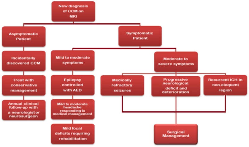
Flow chart showing the management for the symptomatic and asymptomatic cerebral cavernous malformation (CCM) patients. AED indicates antiepileptic drug; and MRI, magnetic resonance imaging.
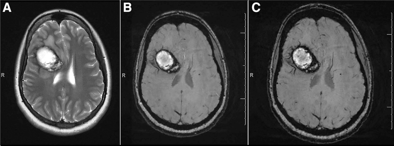
A solitary cerebral cavernous malformation (CCM) with characteristic hypointense ring on (A) T2, (B) T2 *GRE, and (C) SWI. GRE indicates gradient recalled echo; and SWI, susceptibility-weighted imaging.
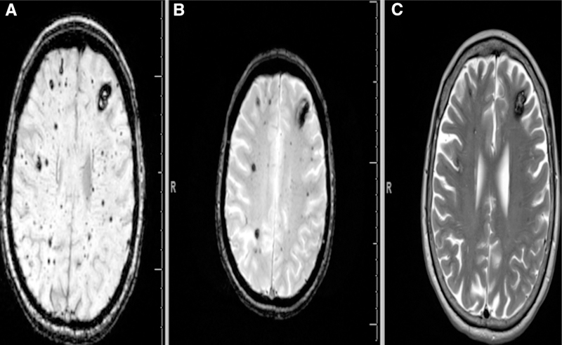
SWI is far more superior to T2*-weighted GRE MRI (B) and conventional T2 sequences (C) to detect smaller type-IV cerebral cavernous malformations (CCMs). Punctate hypointense lesions, black dots with blooming can be noticed both on SWI (A) and T2*-weighted GRE (B). The figure clearly demonstrates SWI to be more sensitive in identifying even small lesions in the same FCCM patient compared with T2*-weighted GRE which can identify most of the cavernomas except micro lesions. FCCM indicates familial cerebral cavernous malformation; GRE, gradient recalled echo; MRI, magnetic resonance imaging; and SWI, susceptibility-weighted imaging.
References
-
- Morrison L, Akers A. Cerebral cavernous malformation, familial In: Adam MP, Ardinger HH, Pagon RA, Wallace SE, Bean LJH, Stephens K, et al., eds. Genereviews® [Internet]. Seattle, WA: University of Washington, Seattle; 1993–2019.
Publication types
MeSH terms
Supplementary concepts
Grants and funding
LinkOut - more resources
Full Text Sources
Medical
