. 2019 Mar 25;38(1):136.
doi: 10.1186/s13046-019-1135-x.

MCUR1 facilitates epithelial-mesenchymal transition and metastasis via the mitochondrial calcium dependent ROS/Nrf2/Notch pathway in hepatocellular carcinoma

Affiliations

MCUR1 facilitates epithelial-mesenchymal transition and metastasis via the mitochondrial calcium dependent ROS/Nrf2/Notch pathway in hepatocellular carcinoma

Mingpeng Jin et al. J Exp Clin Cancer Res. .

Abstract

Background: Mitochondrial Ca2+ plays a critical role in tumorigenesis, including cell proliferation and metastasis. Mitochondrial calcium uniporter regulator 1 (MCUR1) has been shown to be frequently upregulated in HCC and promote cancer cell survival. However, whether MCUR1 is involved in the metastasis of HCC and its underlying mechanisms remain unknown.

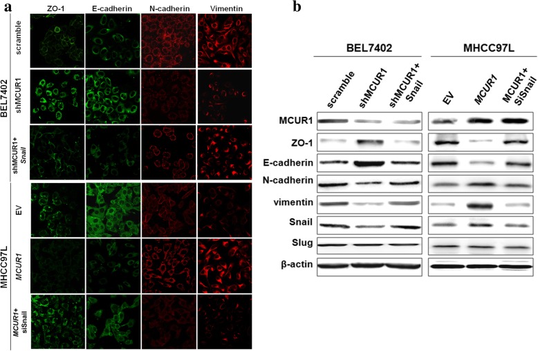
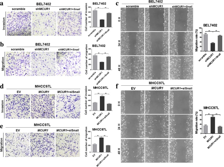
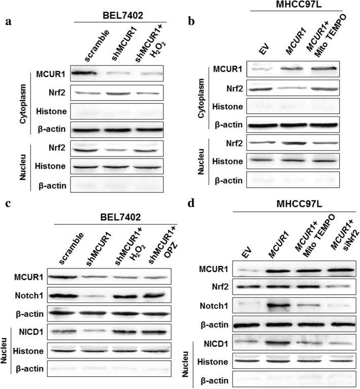
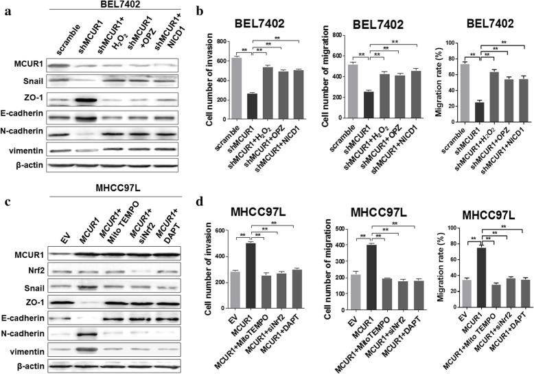
Methods: The effect of MCUR1 expression on epithelial-mesenchymal transition (EMT) in HCC cells was first evaluated by immunofluorescent staining and Western blot. Then, in vitro invasion and in vivo metastasis assays were used to evaluate the function of MCUR1 in HCC metastasis. The underlying mechanism has also been explored by investigating the effect of MCUR1 on ROS/Nrf2/Notch1 pathway.

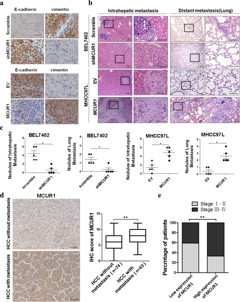
Results: MCUR1 expression was significantly higher in HCC with metastasis and associated with tumor progression. MCUR1 promoted in vitro invasion and in vivo metastasis of HCC cells by promoting EMT via Snail. Mechanistically, MCUR1-mediated mitochondrial Ca2+ signaling promoted the EMT of HCC cells by activating ROS/Nrf2/Notch1 pathway. Inhibition of ROS production, mitochondrial Ca2+ uptake, Nrf2 expression or Notch1 activity significantly suppressed MCUR1-induced EMT of HCC cells. In addition, treatment with the mitochondrial Ca2+-buffering protein parvalbumin significantly inhibited ROS/Nrf2/Notch pathway and MCUR1-induced EMT and HCC metastasis.

Conclusions: Our study provides evidence supporting a metastasis-promoting role for MCUR1-dependent mitochondrial Ca2+ uptake in HCC. Our findings suggest that MCUR1 may be a potential therapeutic target for HCC treatment.

Keywords: EMT; Hepatocellular carcinoma; Metastasis; Mitochondrial calcium uniporter regulator 1; Notch 1.


Conflict of interest statement

Ethics approval and consent to participate

The study was approved by the Ethics Committee of the Fourth Military Medical University. Animal research was approved by the Institutional Animal Care and Use Committee of Fourth Military Medical University. The study was performed in accordance with the Declaration of Helsinki.

Consent for publication

Not applicable.

Competing interests

The authors declare that they have no competing interests.

Publisher’s Note

Springer Nature remains neutral with regard to jurisdictional claims in published maps and institutional affiliations.

Figures

Fig. 1
Fig. 1
MCUR1 promoted EMT of HCC cells. a Immunofluorescent image of epithelial markers (ZO-1 and E-cadherin) and mesenchymal markers (N-cadherin and Vimentin) in HCC cells with different MCUR1 expression levels. Cells were transfected with siRNA or expression vector for 48 h. Scramble: vector encoding control shRNA; shMCUR1 vectors encoding short hairpin RNA (shRNA) against MCUR1. EV: Empty Vector; MCUR1: expression vectors encoding MCUR1; Snail, Snail expression vector; siSnail, siRNA against Snail. b Western blot analysis of ZO-1, E-cadherin, N-cadherin, Vimentin, Snail and Slug in HCC cells with different MCUR1 expression levels
Fig. 2
Fig. 2
MCUR1 promoted in vitro invasion and migration of HCC cells by snail-mediated EMT. Transwell assay for ability of invasion (a, d) and migration (b, e) in stably transfected HCC cells with treatment as indicated. c, f Representative images of wound-healing assay and corresponding statistical analysis in stably transfected HCC cells with treatment as indicated. Data shown are the mean ± SD from three independent experiments. * P < 0.05; ** P < 0.01
Fig. 3
Fig. 3
MCUR1 facilitates epithelial-mesenchymal transition and HCC metastasis. a Representative immunohistochemical staining images of E-cadherin and Vimentin in the orthotopic transplantation nude mice model of HCC metastasis. b Histological analyses of intrahepatic and lung metastatic nodules from HCC metastasis nude mice model by hematoxylin and eosin (H&E) staining. Images showing representative H&E staining of liver and lung tissue samples from the different experimental groups (n = 5 /group). c The number of intrahepatic and lung metastasis nodules was quantified in H.E. sections. d Representative immunohistochemical staining images and IHC score of MCUR1 in HCC without metastasis (n = 74) and HCC with metastasis (n = 63). e MCUR1 expression positively correlated with tumor progression of HCC patients. Data shown are the mean ± SD from three independent experiments. * P < 0.05; ** P < 0.01
Fig. 4
Fig. 4
MCUR1 facilitates ROS-induced Nrf2 nuclear translocation to activate the Notch pathway. a, b, c, d Western blot analysis for expression levels of Nrf2, Notch1and NICD1 in stably transfected HCC cells with treatments as indicated
Fig. 5
Fig. 5
Activation of ROS/Nrf2/Notch pathways is essential for MCUR1-induced EMT and HCC metastasis. a, c Western blot analysis for expression levels of Snail, ZO-1, E-cadherin, N-cadherin and Vimentin in stably transfected HCC cells with treatments as indicated. b, d Transwell assay for migration and invasion ability and wound healing assays for migration rate in stably transfected HCC cells with treatment as indicated. H2O2 (25 μM, for 1 h), OPZ (20 μM, for 1 h), MitoTEMPO (5 μM for 1 h), DAPT (2 μM, for 6 h); cells were transfected with siRNA or expression vector for 48 h. NICD1, NICD1 expression vector; siNrf2, siRNA against Nrf2. Migration rate% = (Migrated distance at indicated time - initial distance) / (initial distance) × 100%. Data shown are the mean ± SD from three independent experiments. * P < 0.05; ** P < 0.01
Fig. 6
Fig. 6
Mitochondrial calcium buffering inhibits ROS/Nrf2/Notch pathway and MCUR1-induced EMT and HCC metastasis. a Western blot analysis for expression levels of Nrf2, Notch1and NICD1 in stably transfected HCC cells with treatments as indicated. Mitochondrial Ca2+ was buffered by transient transfection of expression vector encoding parvalbumin with mitochondria target sequence (PV-Mito) for 48 h, where appropriate. b Western blot analysis for expression levels of ZO-1, E-cadherin, N-cadherin, Vimentin and Snail in stably transfected HCC cells with treatments as indicated. c Transwell assays for migration and invasion ability and wound healing assays for migration rate in stably transfected HCC cells with treatment as indicated. d Representative immunohistochemical staining images of E-cadherin and Vimentin in EV, MCUR1 and the Ad-PV-Mito groups (injection of PV-Mito adenovirus into tail vein). e Histological analyses of intrahepatic and lung metastatic nodules in EV, MCUR1 and the Ad-PV-Mito group (injection of PV-Mito adenovirus into tail vein). Images showing representative H&E staining of liver and lung tissue samples from the different experimental groups (n = 5 /group). f The number of intrahepatic and lung metastasis nodules was quantified in H.E. sections. g The schematic for mechanism underlying epithelial-mesenchymal transition and metastasis facilitated by MCUR1. Data shown are the mean ± SD from three independent experiments. * P < 0.05; ** P < 0.01

Similar articles

Cited by

References

    1. Ferlay J, Soerjomataram I, Dikshit R, Eser S, Mathers C, Rebelo M, Parkin DM, Forman D, Bray F. Cancer incidence and mortality worldwide: sources, methods and major patterns in GLOBOCAN 2012. Int J Cancer. 2015;136(5):E359–E386. doi: 10.1002/ijc.29210. - DOI - PubMed
    1. Giannelli G, Koudelkova P, Dituri F, Mikulits W. Role of epithelial to mesenchymal transition in hepatocellular carcinoma. J Hepatol. 2016;65(4):798–808. doi: 10.1016/j.jhep.2016.05.007. - DOI - PubMed
    1. Van't Veer LJ, Weigelt B. Road map to metastasis. Nat Med. 2003;9(8):999–1000. doi: 10.1038/nm0803-999b. - DOI - PubMed
    1. Budhu A, Forgues M, Ye QH, Jia HL, He P, Zanetti KA, Kammula US, Chen Y, Qin LX, Tang ZY, et al. Prediction of venous metastases, recurrence, and prognosis in hepatocellular carcinoma based on a unique immune response signature of the liver microenvironment. Cancer Cell. 2006;10(2):99–111. doi: 10.1016/j.ccr.2006.06.016. - DOI - PubMed
    1. Prevarskaya N, Skryma R, Shuba Y. Calcium in tumour metastasis: new roles for known actors. Nat Rev Cancer. 2011;11(8):609–618. doi: 10.1038/nrc3105. - DOI - PubMed

MeSH terms