

Transmission of Anaplasma phagocytophilum (Foggie, 1949) by Ixodes ricinus (Linnaeus, 1758) ticks feeding on dogs and artificial membranes
- PMID: 30909972
- PMCID: PMC6434881
- DOI: 10.1186/s13071-019-3396-9
Transmission of Anaplasma phagocytophilum (Foggie, 1949) by Ixodes ricinus (Linnaeus, 1758) ticks feeding on dogs and artificial membranes
Abstract
Background: The interplay of speed of activity of acaricidal products and tick-borne pathogen transmission time is the major driver for disease prevention. This study aimed to investigate the time required for transmission of Anaplasma phagocytophilum by adult Ixodes ricinus ticks in vivo on dogs, and to confirm the time required for transmission observed in vivo, in vitro.
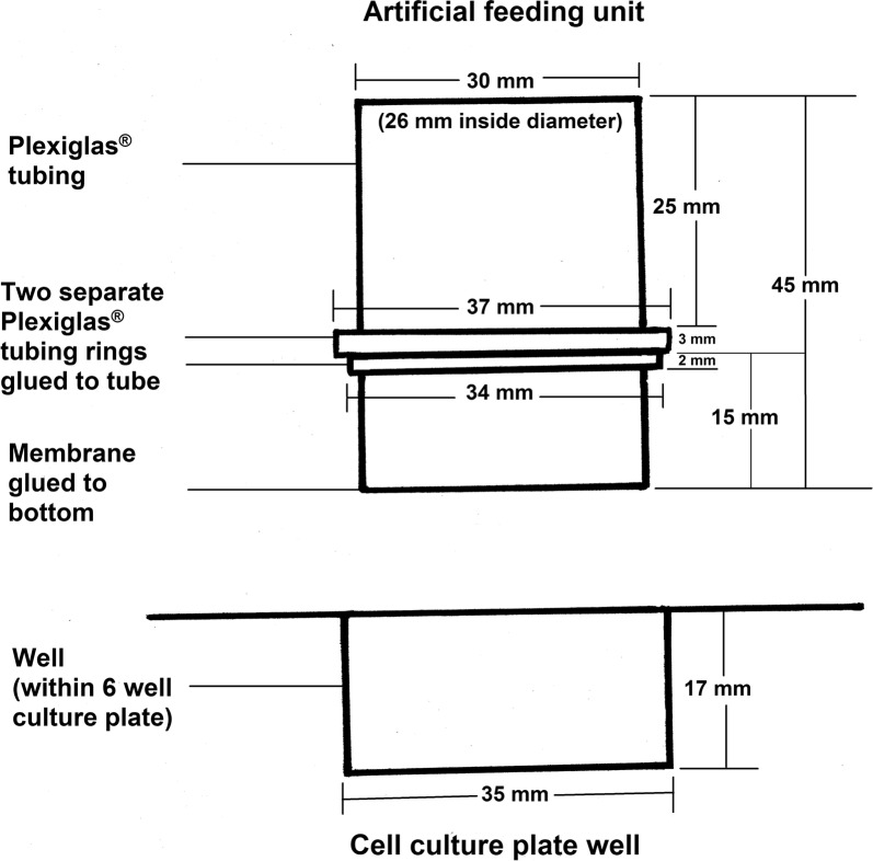
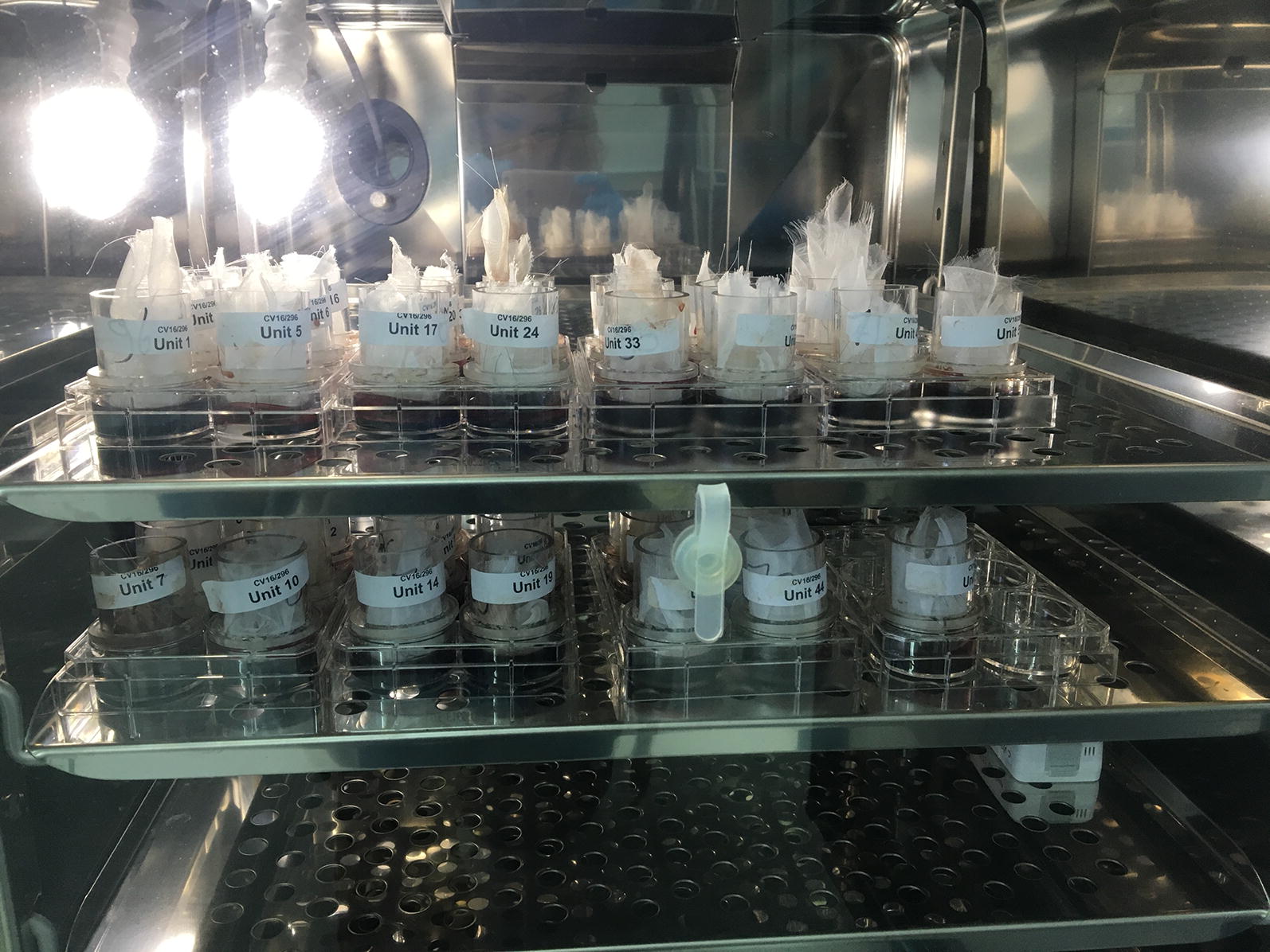
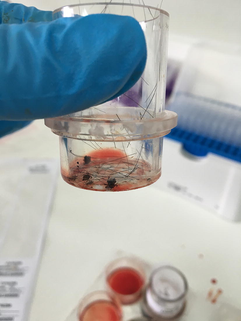
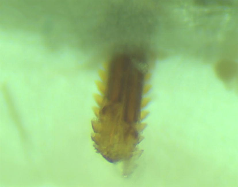
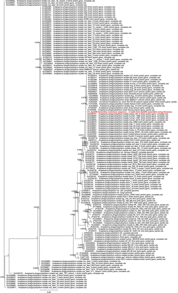
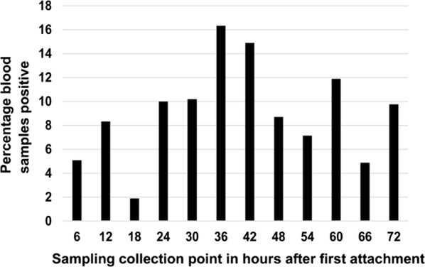
Methods: Nymphs of I. ricinus were experimentally infected with an A. phagocytophilum strain of canine origin. Dogs were allocated to 6 groups of 3 dogs each. Groups 1-5 were infested with 50 A. phagocytophilum-infected female adult ticks on Day 0. Ticks were removed post-infestation at 3, 6, 12, 24 and 48 h. Dogs in Group 6 were infested with 60 A. phagocytophilum-infected female adult ticks (left on dogs until engorged). Dogs were observed daily for general health and clinically examined on Day 0, and weekly from Day 14. Blood was collected for qPCR and serological analysis on Day 0 (pre-challenge) and weekly thereafter. In the in vitro study each artificial feeding chamber was seeded with 10 adult ticks (5 male/5 female), attachment assessed, and blood pools sampled for qPCR at 6 h intervals up to 72 h after first tick attachment.
Results: Anaplasma phagocytophilum specific antibodies and DNA were detected in all 3 dogs in Group 6. No A. phagocytophilum-specific antibodies or DNA were detected in any dogs in Groups 1-5. All dogs remained healthy. Female tick attachment in 60 artificial feeding chambers over 72 h ranged between 20-60%. Anaplasma phagocytophilum DNA was detected in the blood collected from 5% of chambers sampled at 6 h, with the highest number of positive samples (16.3%) observed at 36 h.
Conclusions: Transmission of A. phagocytophilum by I. ricinus ticks starts within a few hours after attachment but establishment of infections in dogs is apparently dependent on a minimum inoculation dose that was only observed when ticks attached for greater than 48 h. These findings highlight the need for acaricidal products to exert a repellent and/or rapid killing effect on ticks to forestall transmission and subsequent disease.
Keywords: Anaplasma phagocytophilum; Dogs; In vitro; In vivo; Ixodes ricinus; Transmission.
Conflict of interest statement
JF and AE are employed by Clinvet and DC, ML and MM by Clinglobal, the organizations that performed the study for Bayer Animal Health (sponsor company who funded the study). MP and BS are employed by Bayer Animal Health GmbH.
Figures
References
MeSH terms
Substances
Grants and funding
LinkOut - more resources
Full Text Sources
Medical
